Copyright
©2011 Baishideng Publishing Group Co.
World J Gastrointest Endosc. May 16, 2011; 3(5): 86-94
Published online May 16, 2011. doi: 10.4253/wjge.v3.i5.86
Published online May 16, 2011. doi: 10.4253/wjge.v3.i5.86
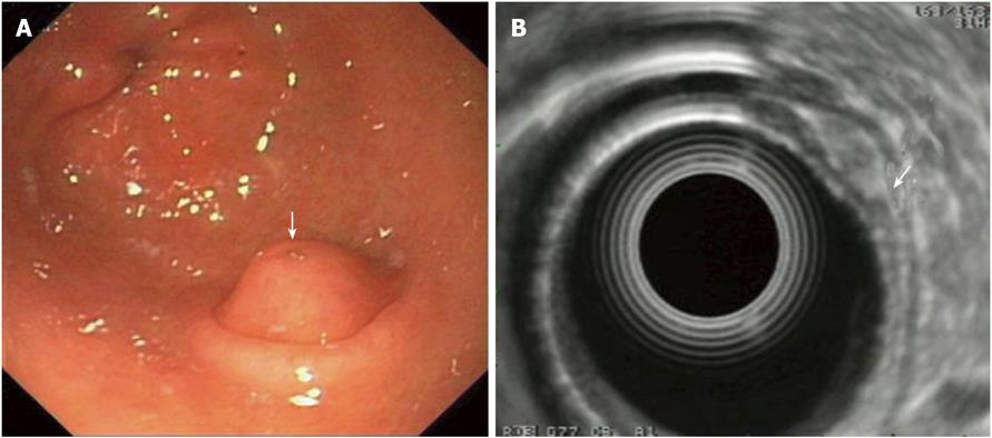
Figure 3 Pancreatic rest of the stomach: Endoscopic and endoscopic ultrasonography -imaging.
A: Endoscopic image of a pancreatic rest. Note the duct opening on the surface of lesion is covered by a normal mucosa with a central umbilication (arrow); B: EUS imaging of the lesion which originates from the 3rd layer, i.e. the submucosa (arrow); note the lesion’s mixed echogenicity. EUS: Endoscopic ultrasonography.
- Citation: Papanikolaou IS, Triantafyllou K, Kourikou A, Rösch T. Endoscopic ultrasonography for gastric submucosal lesions. World J Gastrointest Endosc 2011; 3(5): 86-94
- URL: https://www.wjgnet.com/1948-5190/full/v3/i5/86.htm
- DOI: https://dx.doi.org/10.4253/wjge.v3.i5.86


