
















©2011 Baishideng Publishing Group Co.
World J Gastrointest Endosc. Nov 16, 2011; 3(11): 220-224
Published online Nov 16, 2011. doi: 10.4253/wjge.v3.i11.220
Published online Nov 16, 2011. doi: 10.4253/wjge.v3.i11.220
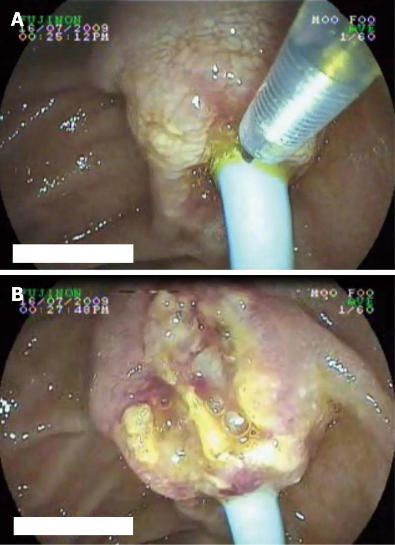
Figure 1 Duodenoscopy showing a periampullary mass with stent in situ and needle knife coming out of endoscope (A) and endoscopic image after needle knife papillotomy showing exposed tumor tissue (B).
- Citation: Noor MT, Vaiphei K, Nagi B, Singh K, Kochhar R. Role of needle knife assisted ampullary biopsy in the diagnosis of periampullary carcinoma. World J Gastrointest Endosc 2011; 3(11): 220-224
- URL: https://www.wjgnet.com/1948-5190/full/v3/i11/220.htm
- DOI: https://dx.doi.org/10.4253/wjge.v3.i11.220