Copyright
©2011 Baishideng Publishing Group Co.
World J Gastrointestinal Endoscopy. Oct 16, 2011; 3(10): 183-194
Published online Oct 16, 2011. doi: 10.4253/wjge.v3.i10.183
Published online Oct 16, 2011. doi: 10.4253/wjge.v3.i10.183
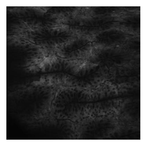
Figure 6 Normal colonic mucosa seen with a confocal endomicroscope.
Glands are seen longitudinally but are well organized and homogenous. Goblet cells appear black.
- Citation: Shukla R, Abidi WM, Richards-Kortum R, Anandasabapathy S. Endoscopic imaging: How far are we from real-time histology? World J Gastrointestinal Endoscopy 2011; 3(10): 183-194
- URL: https://www.wjgnet.com/1948-5190/full/v3/i10/183.htm
- DOI: https://dx.doi.org/10.4253/wjge.v3.i10.183


