
















©2010 Baishideng.
World J Gastrointest Endosc. Apr 16, 2010; 2(4): 121-129
Published online Apr 16, 2010. doi: 10.4253/wjge.v2.i4.121
Published online Apr 16, 2010. doi: 10.4253/wjge.v2.i4.121
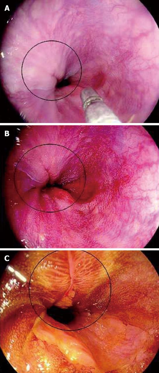
Figure 4 Images from i-SCAN.
A: HD+: A mucosal break is not clearly visible; B: i-SCAN can better depict the mucosal break; C: Lugol’s stain reveals a larger extent of the mucosal break which can help upgrade the degree of esophagitis according to Los Angeles classification. (with permission from Georg Thieme Verlag KG).
- Citation: Chaiteerakij R, Rerknimitr R, Kullavanijaya P. Role of digital chromoendoscopy in detecting minimal change esophageal reflux disease. World J Gastrointest Endosc 2010; 2(4): 121-129
- URL: https://www.wjgnet.com/1948-5190/full/v2/i4/121.htm
- DOI: https://dx.doi.org/10.4253/wjge.v2.i4.121