Copyright
©2010 Baishideng.
World J Gastrointest Endosc. Mar 16, 2010; 2(3): 90-96
Published online Mar 16, 2010. doi: 10.4253/wjge.v2.i3.90
Published online Mar 16, 2010. doi: 10.4253/wjge.v2.i3.90
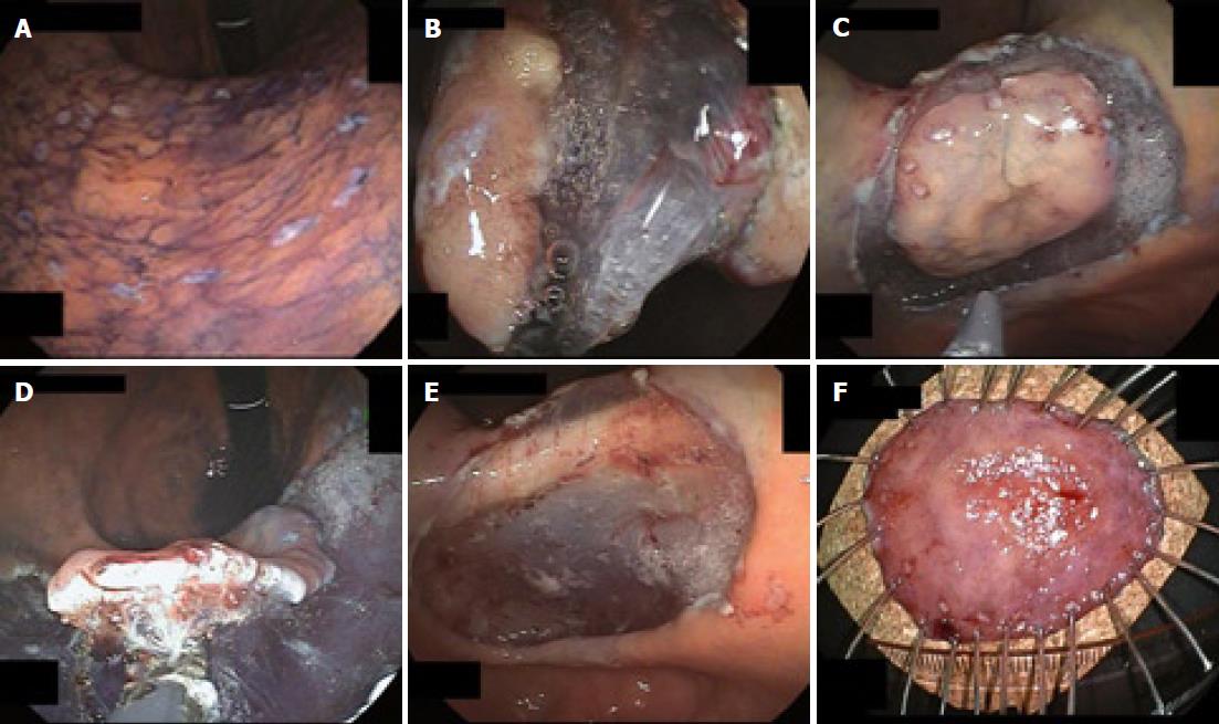
Figure 4 Endoscopic view of the procedure of ESD using GSF.
A: Marks are made at several points along the outline of the lesion with a coagulation current; B: The mucosa is incised outside the marker dots to separate the lesion from the surrounding non-neoplastic mucosa using GSF; C: Completion of the GSF cutting around the lesion with a safe lateral margin; D: The submucosal connective tissue beneath the lesion is grasped and lifted up and excised using GSF from the underlying muscle layer; E: The lesion is cut completely from the muscle layer; F: The resected specimen showing en bloc resection of the lesion.
- Citation: Akahoshi K, Akahane H. A new breakthrough: ESD using a newly developed grasping type scissor forceps for early gastrointestinal tract neoplasms. World J Gastrointest Endosc 2010; 2(3): 90-96
- URL: https://www.wjgnet.com/1948-5190/full/v2/i3/90.htm
- DOI: https://dx.doi.org/10.4253/wjge.v2.i3.90


