Copyright
©2010 Baishideng.
World J Gastrointest Endosc. Feb 16, 2010; 2(2): 69-74
Published online Feb 16, 2010. doi: 10.4253/wjge.v2.i2.69
Published online Feb 16, 2010. doi: 10.4253/wjge.v2.i2.69
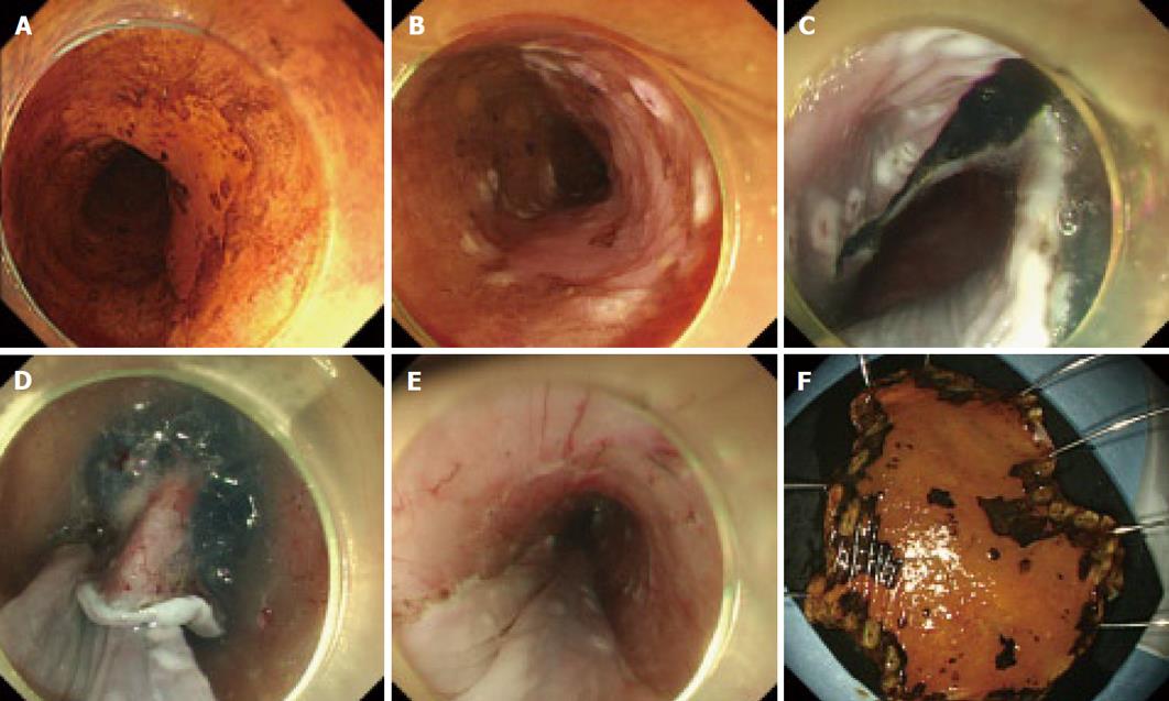
Figure 1 Typical example case of ESD for esophageal neoplasm.
A: Chromoendoscopy with iodine staining to demarcate the lesion from the non-neoplastic area; B: Marking placement around the lesion; C: Initial mucosal incision after submucosal injection at the distal margin of the lesions; D: Mucosal incision after submucosal injection at the proximal margin of the lesion and subsequent submucosal dissection from the proximal end; E: Mucosal defect after ESD; F: Resected specimen.
- Citation: Nonaka K, Arai S, Ishikawa K, Nakao M, Nakai Y, Togawa O, Nagata K, Shimizu M, Sasaki Y, Kita H. Short term results of endoscopic submucosal dissection in superficial esophageal squamous cell neoplasms. World J Gastrointest Endosc 2010; 2(2): 69-74
- URL: https://www.wjgnet.com/1948-5190/full/v2/i2/69.htm
- DOI: https://dx.doi.org/10.4253/wjge.v2.i2.69


