
















Copyright: ©Author(s) 2026.
World J Gastrointest Endosc. Aug 16, 2026; 18(8): 122701
Published online Aug 16, 2026. doi: 10.4253/wjge.122701
Published online Aug 16, 2026. doi: 10.4253/wjge.122701
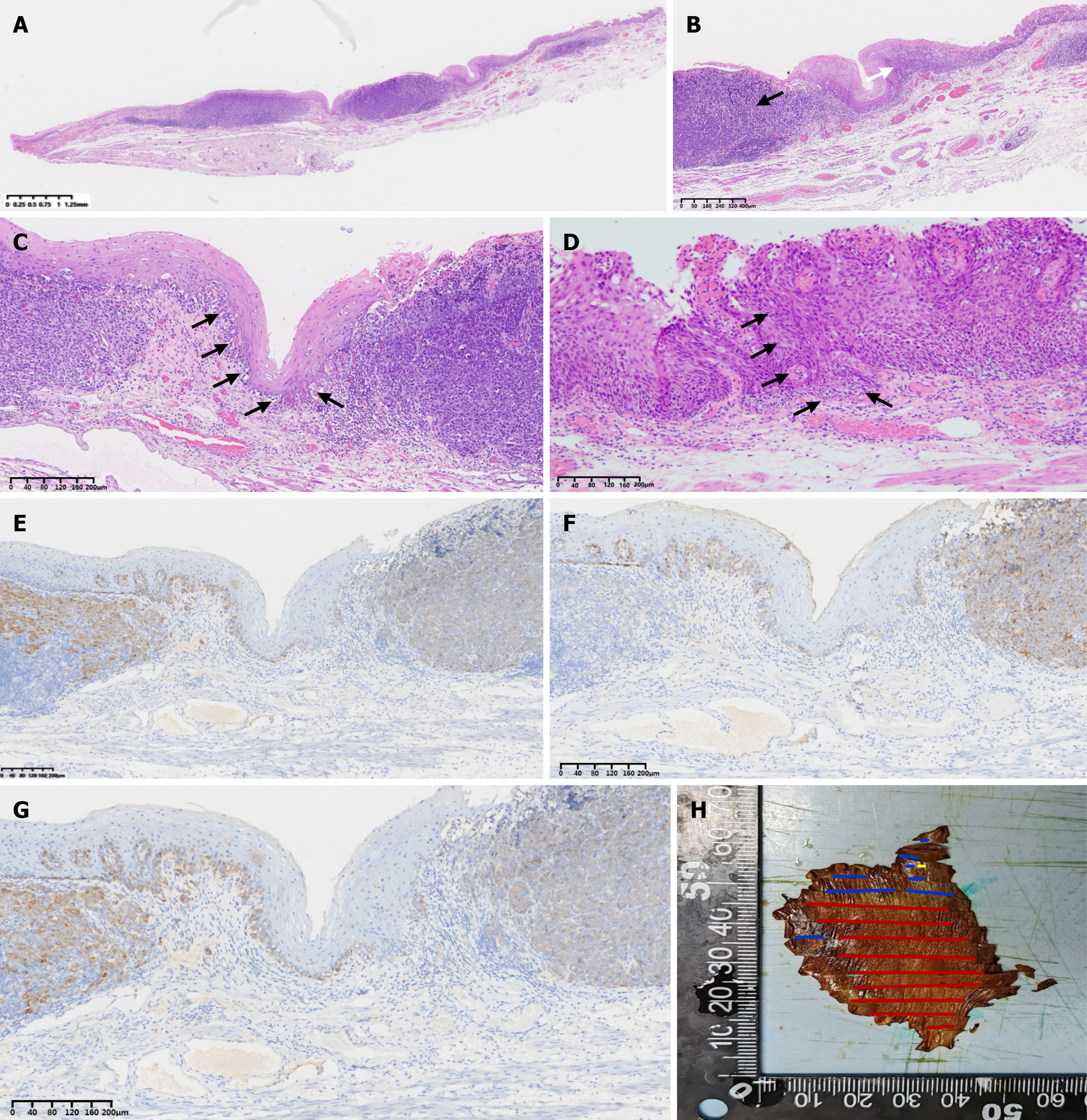
Figure 2 Histological and immunohistochemical findings of the collision tumor.
A: Tumor cells are separated from the resection margins by a safety distance of > 1 mm (H&E, × 10); B: Low-power view showing the overall architecture of the collision tumor (H&E, × 40). The dashed line demarcates the boundary between high-grade intraepithelial neoplasia (right) and the primary malignant melanoma of the esophagus (left); C: Higher magnification of the adjacent squamous epithelium revealing melanocytic hyperplasia in the basal layer, consistent with junctional activity (H&E, × 200); D: An adjacent section of the same specimen reveals a distinct focus of invasive squamous cell carcinoma that has broken through the basement membrane, fulfilling the diagnostic criteria for squamous cell carcinoma (H&E, × 200); E: Immunohistochemistry showing diffuse positivity for Melan-A in melanoma cells (× 200); F: Immunohistochemistry showing diffuse positivity for HMB45 in melanoma cells (× 200); G: Immunohistochemistry showing diffuse positivity for S-100 protein in melanoma cells (× 200); H: Reconstructed pathological sample maps showing the spatial distribution of the primary malignant melanoma of the esophagus (yellow line), high-grade intraepithelial neoplasia (blue line), and squamous cell carcinoma (red line).
- Citation: Hao YY, Cui JF, Ji CG, Liu L, Yin KG. Endoscopic submucosal dissection for malignant melanoma and esophageal squamous cell carcinoma: A case report and review of literature. World J Gastrointest Endosc 2026; 18(8): 122701
- URL: https://www.wjgnet.com/1948-5190/full/v18/i8/122701.htm
- DOI: https://dx.doi.org/10.4253/wjge.122701