
















Copyright: ©Author(s) 2026.
World J Gastrointest Endosc. Aug 16, 2026; 18(8): 118395
Published online Aug 16, 2026. doi: 10.4253/wjge.118395
Published online Aug 16, 2026. doi: 10.4253/wjge.118395
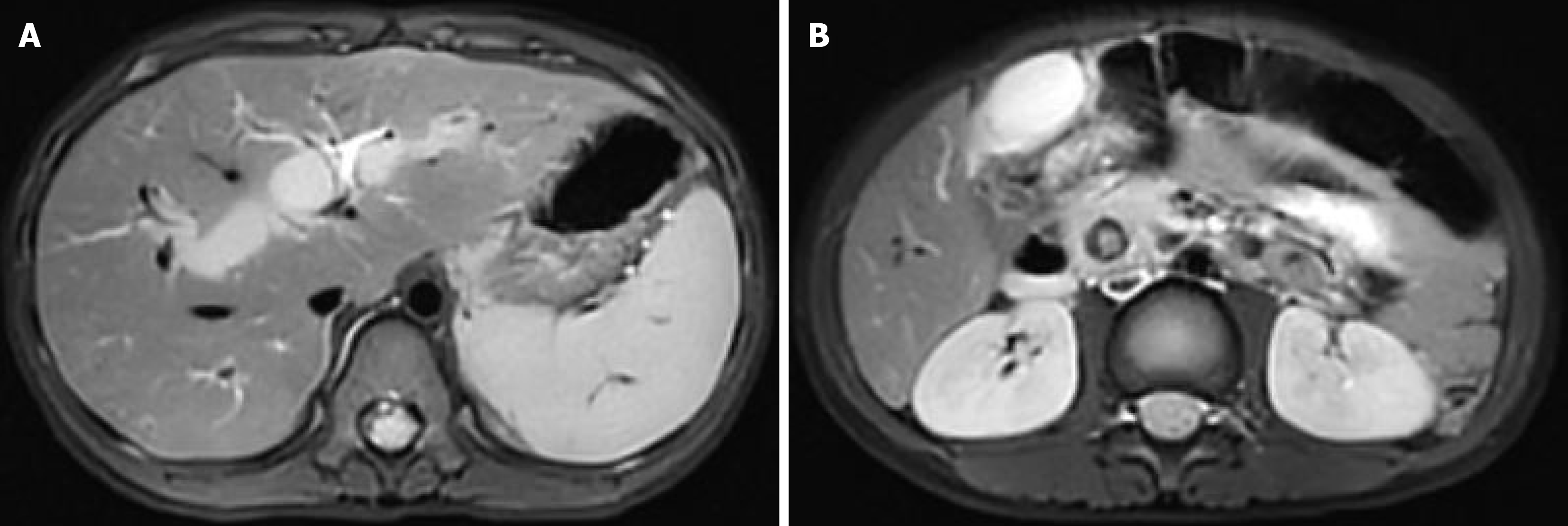
Figure 2 Preoperative abdominal computed tomography findings of common bile duct stones.
A: Coronal computed tomography image demonstrating intrahepatic bile duct dilatation; B: Axial computed tomography image showing a calculus within the common bile duct.
- Citation: Wang YS, Hou M, Li ZY, Du ZJ, Liu JY. Therapeutic endoscopic retrograde cholangiopancreatography for giant bile duct stones in a child: A case report. World J Gastrointest Endosc 2026; 18(8): 118395
- URL: https://www.wjgnet.com/1948-5190/full/v18/i8/118395.htm
- DOI: https://dx.doi.org/10.4253/wjge.118395