
















Copyright: ©Author(s) 2026.
World J Gastrointest Endosc. May 16, 2026; 18(5): 119573
Published online May 16, 2026. doi: 10.4253/wjge.v18.i5.119573
Published online May 16, 2026. doi: 10.4253/wjge.v18.i5.119573
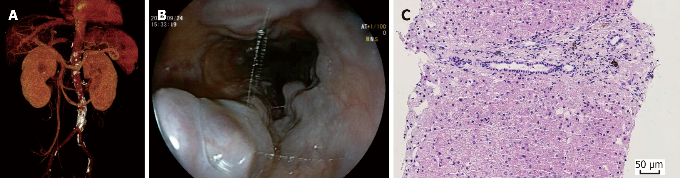
Figure 1 Absence of the hepatic segment of the inferior vena cava complicated by noncirrhotic portal hypertension with esophagogastric varices.
A: Three-dimensional volume-rendered computed tomography reconstruction demonstration of the absence of the hepatic segment of the inferior vena cava with azygos continuation; B: Esophagogastroduodenography showed large proximal esophageal varices with positive red wale signs; C: The liver pathological feature revealed moderate fibrosis in portal areas with absent portal vein lumens, and no evidence of perisinusoidal fibrosis or hepatic veno-occlusive disease.
- Citation: Li M, Zhang LK, Qu L, Liu JH, Liu JJ, Sun SZ, Zhang D, Hao NB, Li CZ. Absent inferior vena cava combined with idiopathic noncirrhotic portal hypertension resulting esophagogastric variceal bleeding: A case report. World J Gastrointest Endosc 2026; 18(5): 119573
- URL: https://www.wjgnet.com/1948-5190/full/v18/i5/119573.htm
- DOI: https://dx.doi.org/10.4253/wjge.v18.i5.119573