
















Copyright: ©Author(s) 2026.
World J Gastrointest Endosc. Apr 16, 2026; 18(4): 118485
Published online Apr 16, 2026. doi: 10.4253/wjge.v18.i4.118485
Published online Apr 16, 2026. doi: 10.4253/wjge.v18.i4.118485
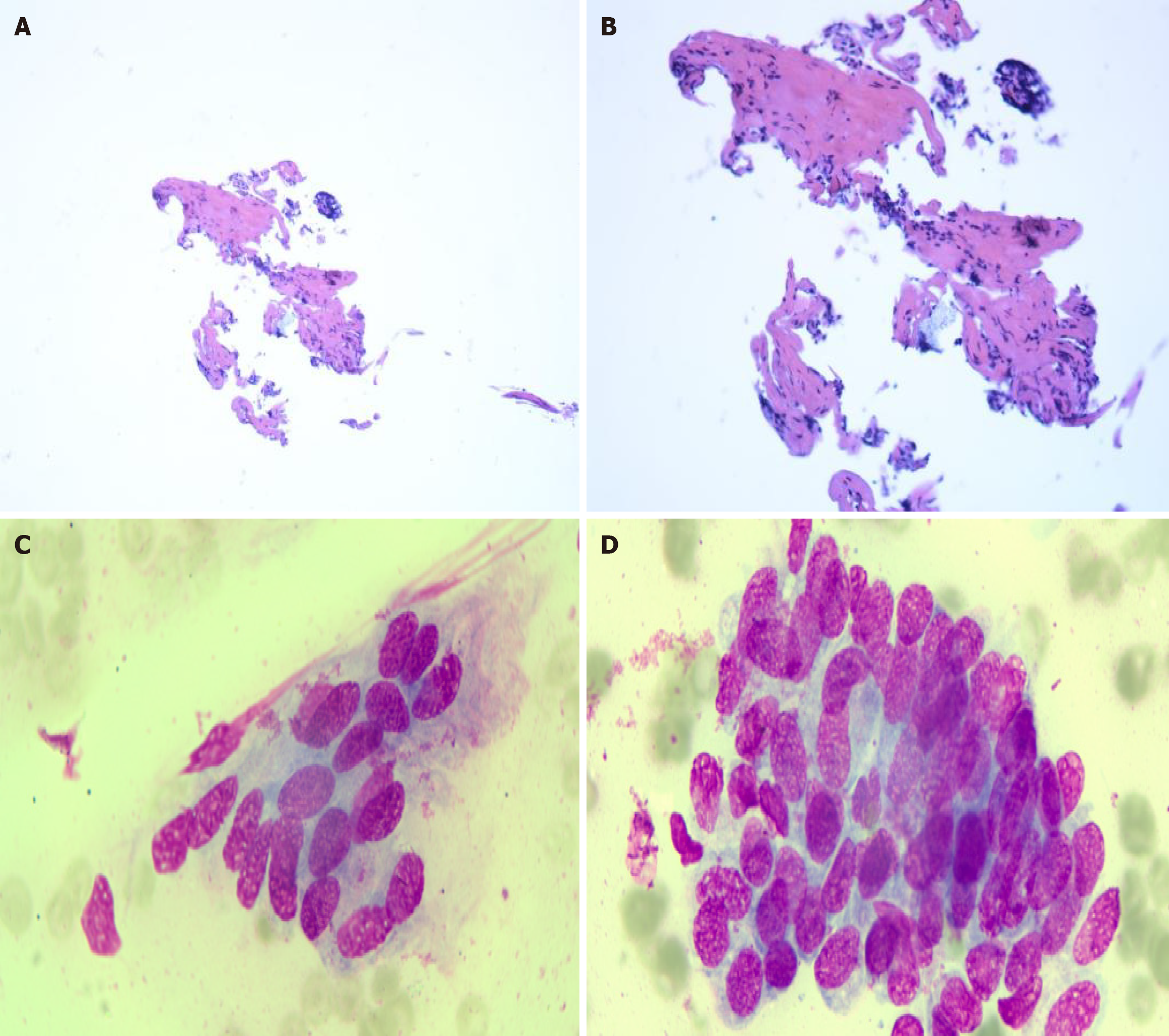
Figure 5 Histopathology and cytopathology.
A and B: Sections of the common bile duct extending to the hepatic bifurcation show minimal fibrosis with chronic inflammatory cell infiltration (A: Hematoxylin and eosin staining, magnification × 40; B: Hematoxylin and eosin staining, magnification × 100); C and D: Bile duct brushings reveal epithelial cells with focal nuclear enlargement, an increased nuclear-to-cytoplasmic ratio, coarse hyperchromatic chromatin, and mild atypia. Correlation with clinical, radiologic, and histologic data is recommended (Wright-Giemsa staining, magnification × 1000).
- Citation: Xia Y, Zhu JD, Su T, Shi L, Zhao SL, Xu HW, Xu CQ. Immunoglobulin G4-related sclerosing cholangitis: A case report. World J Gastrointest Endosc 2026; 18(4): 118485
- URL: https://www.wjgnet.com/1948-5190/full/v18/i4/118485.htm
- DOI: https://dx.doi.org/10.4253/wjge.v18.i4.118485