
















Copyright: ©Author(s) 2026.
World J Gastrointest Endosc. Apr 16, 2026; 18(4): 118422
Published online Apr 16, 2026. doi: 10.4253/wjge.v18.i4.118422
Published online Apr 16, 2026. doi: 10.4253/wjge.v18.i4.118422
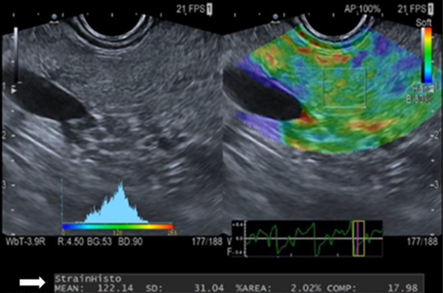
Figure 1 Example image of strain histogram elastography measurement.
A B-mode image is always displayed on the left, and a color-scale elastographic image is superimposed on the right. The region of interest is placed on the pancreatic tissue, and the strain histogram value, resulting from the average of hue histogram values, is shown below (white arrow).
- Citation: Laurenza C, Zignani N, Elvo B, Pignata L, Frigo F, Di Luna I, Soro S, Dagani D, Drago A, Grassia R. Reference range of pancreatic strain histogram by endoscopic ultrasound elastography: A pilot study. World J Gastrointest Endosc 2026; 18(4): 118422
- URL: https://www.wjgnet.com/1948-5190/full/v18/i4/118422.htm
- DOI: https://dx.doi.org/10.4253/wjge.v18.i4.118422