
















Copyright: ©Author(s) 2026.
World J Gastrointest Endosc. Apr 16, 2026; 18(4): 117233
Published online Apr 16, 2026. doi: 10.4253/wjge.v18.i4.117233
Published online Apr 16, 2026. doi: 10.4253/wjge.v18.i4.117233
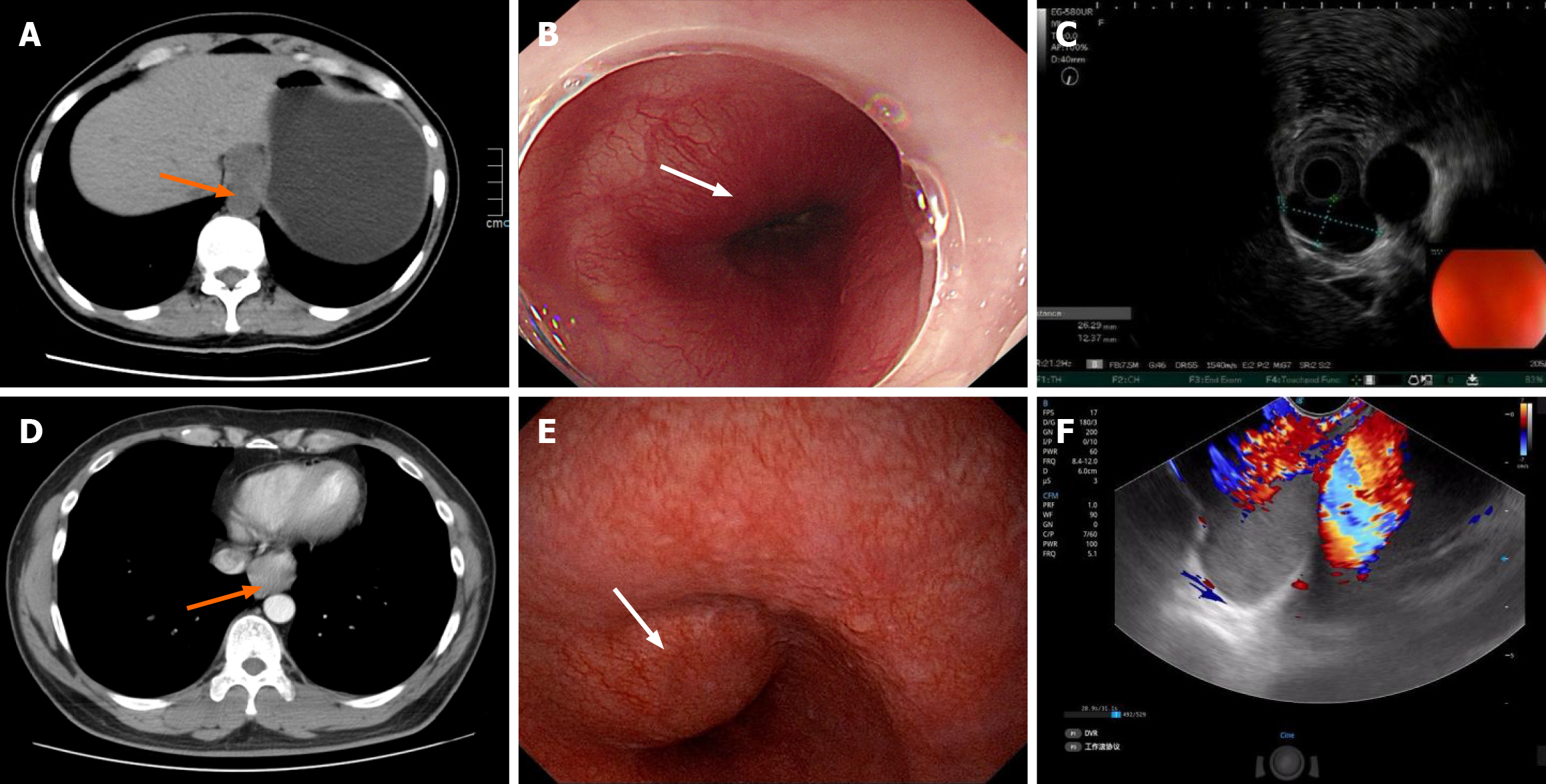
Figure 1 Preoperative imaging and endoscopic findings.
A: Computed tomography shows a well-circumscribed, low-density mass at the gastroesophageal junction (case 1) (arrow indicate the lesions); B: Endoscopy reveals a smooth submucosal bulge 35 cm from the incisors (case 1) (arrow indicate the lesions); C: Endoscopic ultrasonography demonstrates a hypoechoic, irregularly shaped lesion arising from the muscularis propria with clear margins (case 1); D: Contrast-enhanced computed tomography shows a mass adjacent to the gastroesophageal junction with mild enhancement and luminal narrowing (case 2) (arrow indicate the lesions); E: Endoscopy revealing a smooth, intact submucosal bulge 38 cm from the incisors (case 2) (arrow indicate the lesions); F: Endoscopic ultrasonography showing a hypoechoic lesion originating from the muscularis propria, with no vascular signal on color Doppler flow imaging and intermediate stiffness on elastography (case 2).
- Citation: Ma JM, Ji CG, Liu L, Yin KG. Bronchogenic cysts treated with submucosal tunneling endoscopic resection and natural orifice transluminal endoscopy: Two case reports and review of literature. World J Gastrointest Endosc 2026; 18(4): 117233
- URL: https://www.wjgnet.com/1948-5190/full/v18/i4/117233.htm
- DOI: https://dx.doi.org/10.4253/wjge.v18.i4.117233