
















Copyright: ©Author(s) 2026.
World J Gastrointest Endosc. Apr 16, 2026; 18(4): 115366
Published online Apr 16, 2026. doi: 10.4253/wjge.v18.i4.115366
Published online Apr 16, 2026. doi: 10.4253/wjge.v18.i4.115366
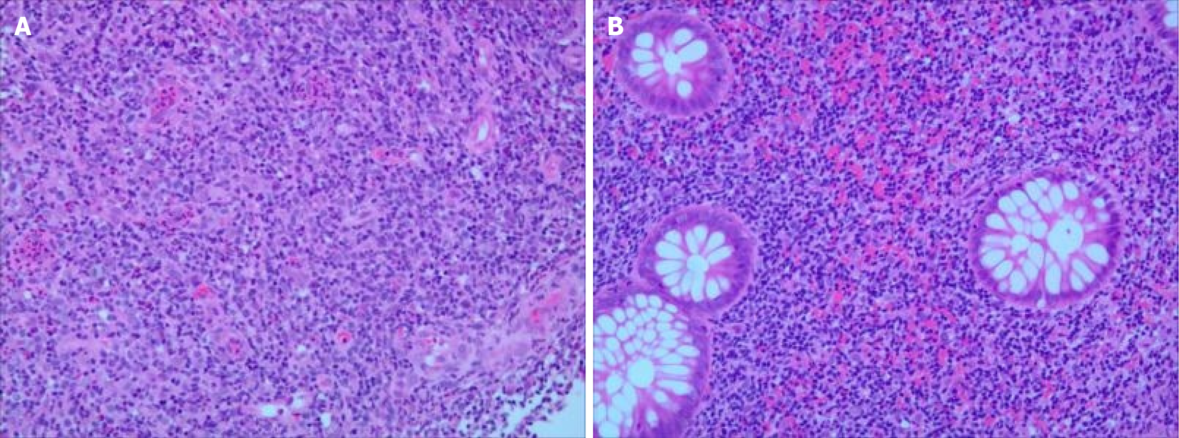
Figure 4 Tissue biopsy pathology findings.
A and B: The tissues (terminal ileum, cecum, ascending colon, transverse colon, descending colon, sigmoid colon, and rectum) were covered with columnar epithelium, the glands were regularly arranged, the cells were not heterogeneous, and the interstitium was infiltrated by many lymphocytes, with small-to-medium sized heterogeneous cells between, fine nuclear chromatin, and inconspicuous nuclei. The foci were clonal proliferation, with low cytoplasmic volume.
- Citation: Yu YM, Huang J, Chen Y, Lu BY. Fecal microbiota transplantation for indolent T-cell lymphoproliferative disorder of gastrointestinal tract: A case report and review of literature. World J Gastrointest Endosc 2026; 18(4): 115366
- URL: https://www.wjgnet.com/1948-5190/full/v18/i4/115366.htm
- DOI: https://dx.doi.org/10.4253/wjge.v18.i4.115366