
















Copyright: ©Author(s) 2026.
World J Gastrointest Endosc. Apr 16, 2026; 18(4): 115366
Published online Apr 16, 2026. doi: 10.4253/wjge.v18.i4.115366
Published online Apr 16, 2026. doi: 10.4253/wjge.v18.i4.115366
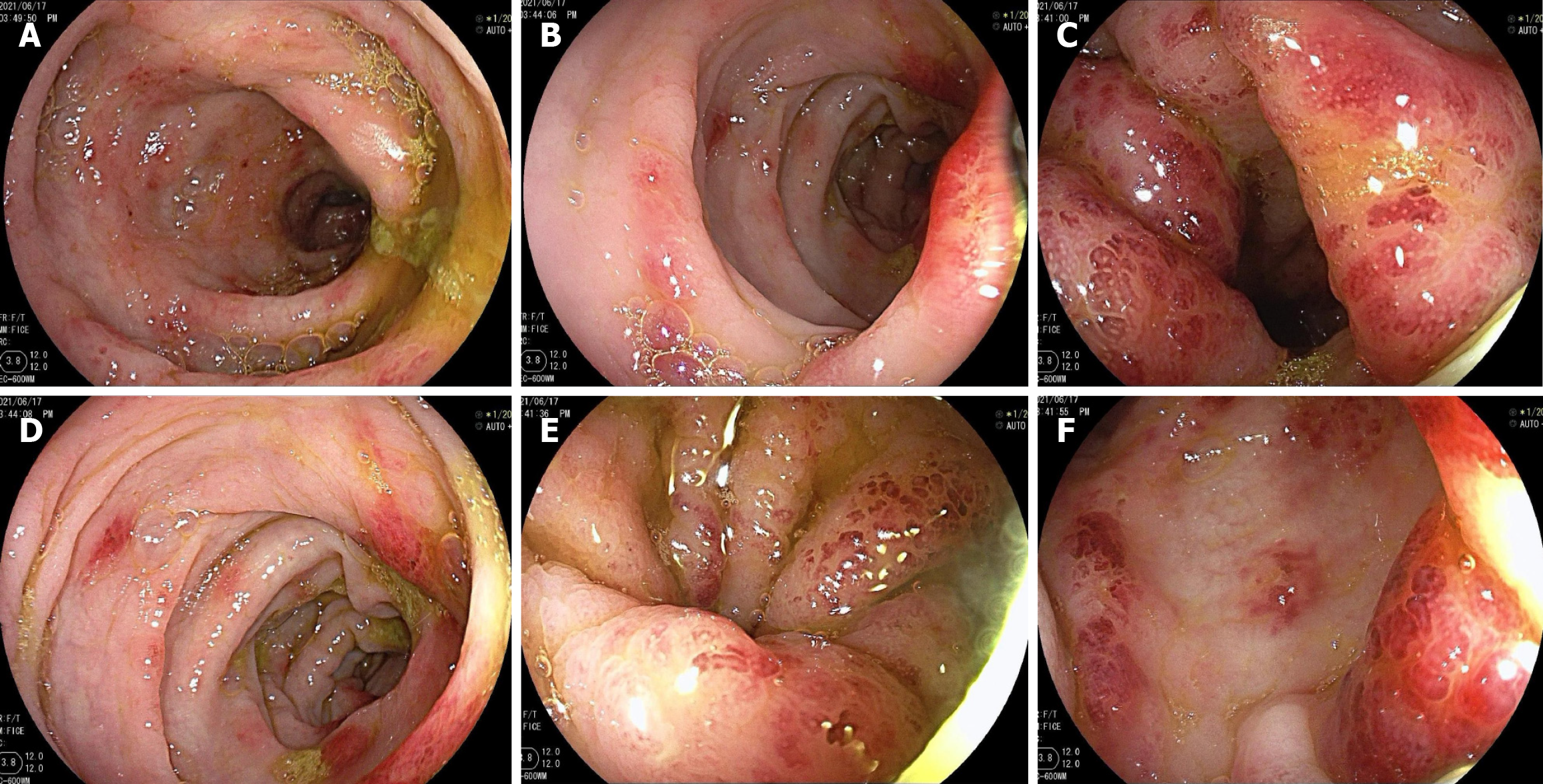
Figure 3 Enteroscopic presentation at admission.
Multiple ulcerated erosions, surface redness, partial white moss covering, extensive intestinal mucosa erosion, congestion, and local oozing blood. The colonic pouch had disappeared, and the intestinal tubes showed regular lead-pipe-like morphology. The half of the colon was more serious, segmental biopsy, tissue elasticity was poor, easy to bleed. A: Ascending colon; B: Transverse colon; C: Descending colon; D: Sigmoid colon; E and F: Rectum.
- Citation: Yu YM, Huang J, Chen Y, Lu BY. Fecal microbiota transplantation for indolent T-cell lymphoproliferative disorder of gastrointestinal tract: A case report and review of literature. World J Gastrointest Endosc 2026; 18(4): 115366
- URL: https://www.wjgnet.com/1948-5190/full/v18/i4/115366.htm
- DOI: https://dx.doi.org/10.4253/wjge.v18.i4.115366