
















Copyright: ©Author(s) 2026.
World J Gastrointest Endosc. Apr 16, 2026; 18(4): 115366
Published online Apr 16, 2026. doi: 10.4253/wjge.v18.i4.115366
Published online Apr 16, 2026. doi: 10.4253/wjge.v18.i4.115366
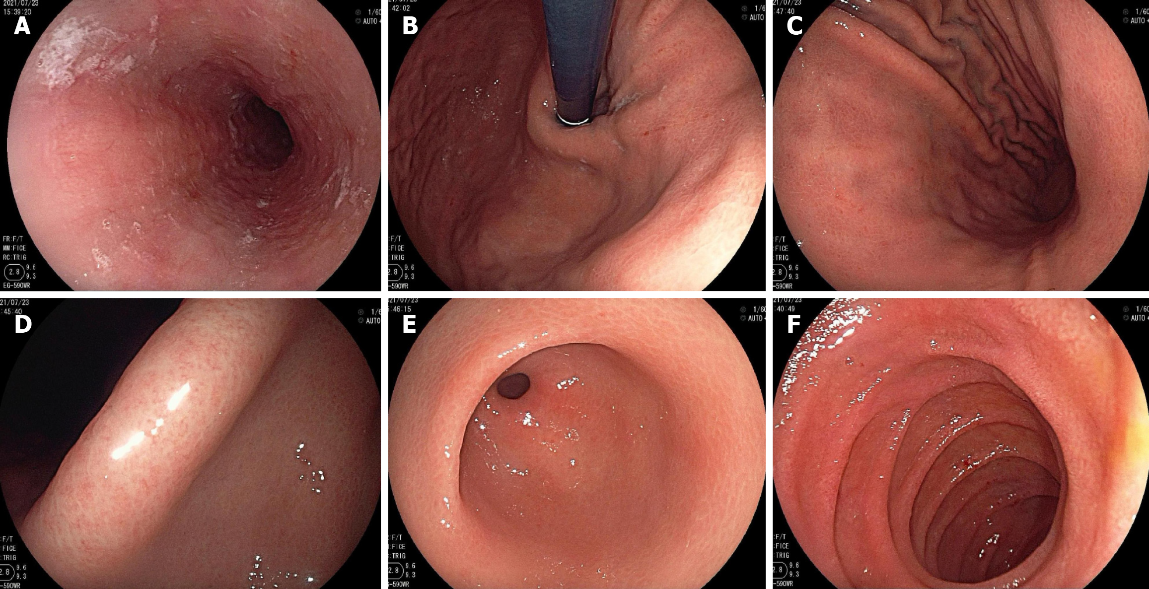
Figure 2 Gastroscopic presentation on admission.
A: The esophagus had a smooth mucosa with a clear vascular network, and no ulcers, erosions, polyps, or varicose veins were seen; B and C: The gastric fundus and the gastric body had multiple red spots, and the regular arrangement of collecting venules was not visible. The mucosa of the gastric body was mildly reddened and swollen without chicken-skin-like changes; D and E: The mucosal regular arrangement of collecting venules was not visible in the gastric angle. The gastric angle and antrum had no chicken-skin-like changes; F: The proximal mucosa and annular folds of the descending duodenum were normal.
- Citation: Yu YM, Huang J, Chen Y, Lu BY. Fecal microbiota transplantation for indolent T-cell lymphoproliferative disorder of gastrointestinal tract: A case report and review of literature. World J Gastrointest Endosc 2026; 18(4): 115366
- URL: https://www.wjgnet.com/1948-5190/full/v18/i4/115366.htm
- DOI: https://dx.doi.org/10.4253/wjge.v18.i4.115366