
















Copyright: ©Author(s) 2026.
World J Gastrointest Endosc. Mar 16, 2026; 18(3): 117303
Published online Mar 16, 2026. doi: 10.4253/wjge.v18.i3.117303
Published online Mar 16, 2026. doi: 10.4253/wjge.v18.i3.117303
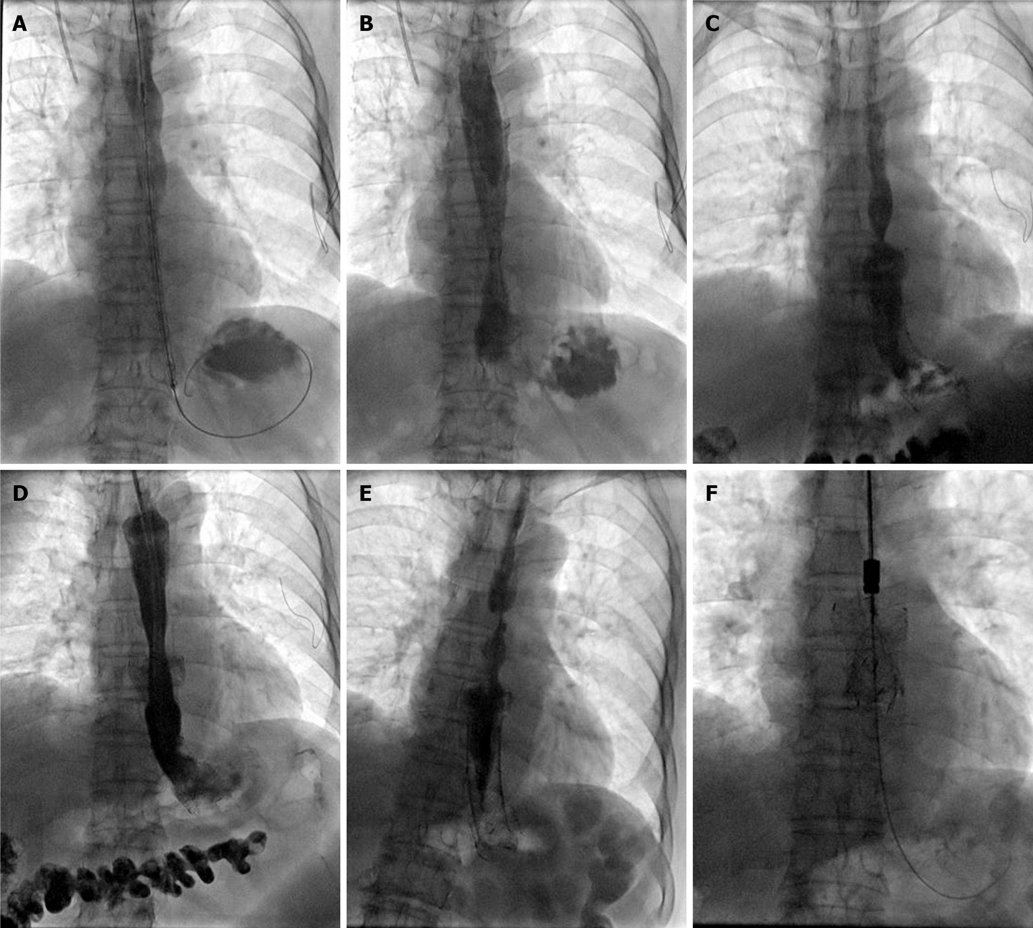
Figure 4 Fluoroscopically guided stenting procedure for the senile patient.
A: A 20 mm × 120 mm oesophageal stent was introduced and inserted in the lower esophagus; B: The oesophagogram immediately after the procedure shows improved passage through the widened lumen of the lower esophagus; C: Restenosis was observed at the proximal end of the stent 2.5 months after stent placement; D: A second 20 mm × 100 mm oesophageal stent was implanted at the stricture site; E: The second stent migrated distally about 2 months after placement, leading to symptom recurrence; F: The two stents were successfully removed under fluoroscopy.
- Citation: Yuan HF, Liu P, Guo CQ, Bi YH. Comparative study of self-expandable stent placement, bougie dilation, and balloon dilation for post-inflammatory oesophageal strictures. World J Gastrointest Endosc 2026; 18(3): 117303
- URL: https://www.wjgnet.com/1948-5190/full/v18/i3/117303.htm
- DOI: https://dx.doi.org/10.4253/wjge.v18.i3.117303