
















Copyright: ©Author(s) 2026.
World J Gastrointest Endosc. Mar 16, 2026; 18(3): 113096
Published online Mar 16, 2026. doi: 10.4253/wjge.v18.i3.113096
Published online Mar 16, 2026. doi: 10.4253/wjge.v18.i3.113096
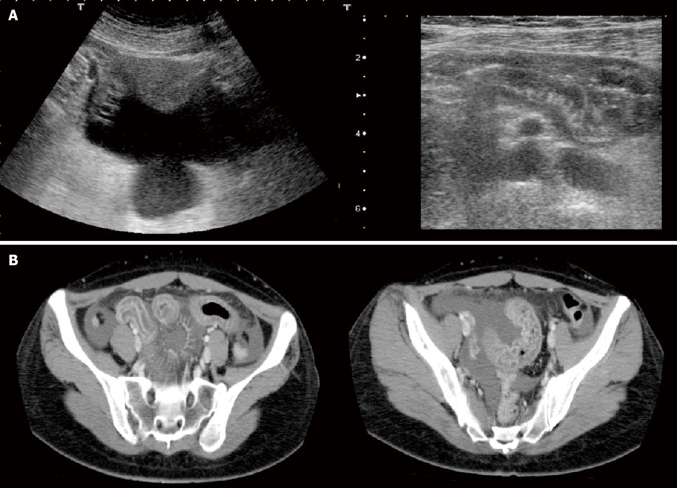
Figure 1 Imaging examinations.
A: Abdominal ultrasound showing significant edema of the pelvic small intestine and ascending colon, and a moderate amount of ascites; B: Abdominal computed tomography scan showing significant edema of the pelvic small intestine and ascending colon, and a moderate amount of ascites.
- Citation: Matsuura K, Ueda C, Hashimura C, Yakushiji H, Horiuchi T. Hereditary angioedema with recurrent abdominal pain in a patient with a novel SERPING1 gene mutation: A case report. World J Gastrointest Endosc 2026; 18(3): 113096
- URL: https://www.wjgnet.com/1948-5190/full/v18/i3/113096.htm
- DOI: https://dx.doi.org/10.4253/wjge.v18.i3.113096