
















©The Author(s) 2026.
World J Gastrointest Endosc. Feb 16, 2026; 18(2): 114400
Published online Feb 16, 2026. doi: 10.4253/wjge.v18.i2.114400
Published online Feb 16, 2026. doi: 10.4253/wjge.v18.i2.114400
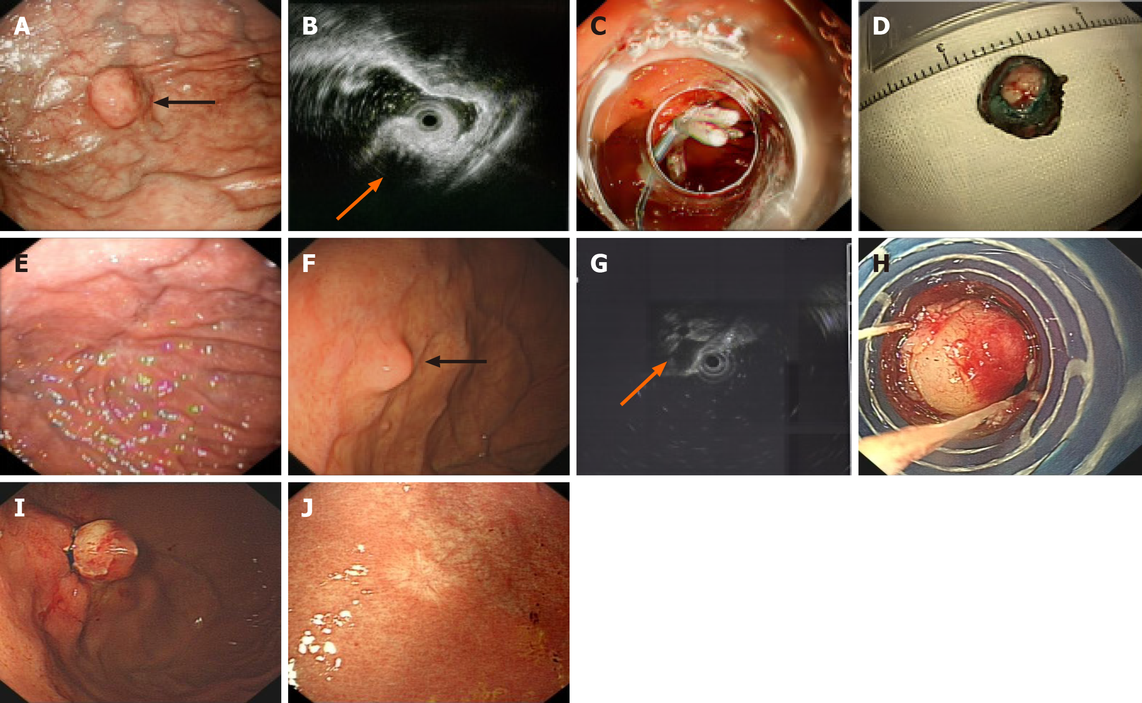
Figure 3 Procedure for endoscopic submucosal dissection and precutting endoscopic band ligation.
A: Preoperative endoscopic view of submucosal tumors originating from the muscularis propria (black arrow); B: Endoscopic ultrasonography view (orange arrow); C: The “clipping closure” procedure in endoscopic submucosal dissection; D: Specimen resected by endoscopic submucosal dissection; E: Five-year endoscopic follow-up after endoscopic submucosal dissection under standard endoscopy; F: Preoperative endoscopic view (black arrow); G: A hypoechoic submucosal tumors originating from the muscularis propria in the gastric fundus (orange arrow) detected by endoscopic ultrasonography; H: The exposed tumor was fully aspirated into the cap; I: The exposed tumor was ligated by band ligation; J: Endoscopic follow-up.
- Citation: Ou Y, Wang J, Liu MF, Yuan R, Min J, Li S, Deng L. Retrospective comparison of precutting endoscopic band ligation and endoscopic submucosal dissection for small gastric muscularis propria tumors. World J Gastrointest Endosc 2026; 18(2): 114400
- URL: https://www.wjgnet.com/1948-5190/full/v18/i2/114400.htm
- DOI: https://dx.doi.org/10.4253/wjge.v18.i2.114400