
















©The Author(s) 2026.
World J Gastrointest Endosc. Feb 16, 2026; 18(2): 113439
Published online Feb 16, 2026. doi: 10.4253/wjge.v18.i2.113439
Published online Feb 16, 2026. doi: 10.4253/wjge.v18.i2.113439
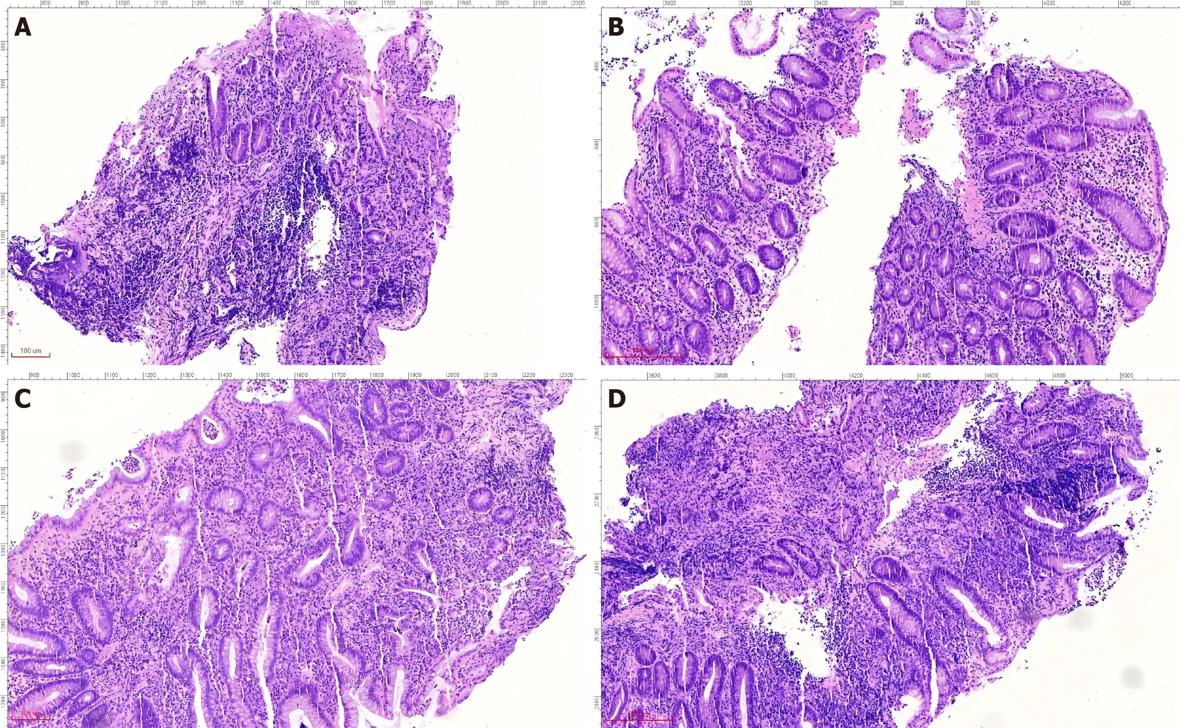
Figure 3 Histopathological diagnosis of ulcerative colitis across multiple colonic sites.
A: Appendix; B: Ileocecal region; C: Sigmoid colon; D: Rectum. The pathological findings of appendiceal, ileocecal, sigmoid, and rectal mucosal biopsies were consistent with ulcerative changes: Uneven distribution of crypts, crypt distortion, visible cryptitis, focal crypt loss, massive plasma cell and lymphocyte infiltration in the stroma, and focal neutrophil infiltration.
- Citation: Wu XJ, Xiang RY, Gong P, Chen Q, Li XF. Relationship between the appendix and ulcerative colitis: A new discovery. World J Gastrointest Endosc 2026; 18(2): 113439
- URL: https://www.wjgnet.com/1948-5190/full/v18/i2/113439.htm
- DOI: https://dx.doi.org/10.4253/wjge.v18.i2.113439