
















Copyright: ©Author(s) 2026.
World J Gastrointest Endosc. Nov 16, 2026; 18(11): 126966
Published online Nov 16, 2026. doi: 10.4253/wjge.126966
Published online Nov 16, 2026. doi: 10.4253/wjge.126966
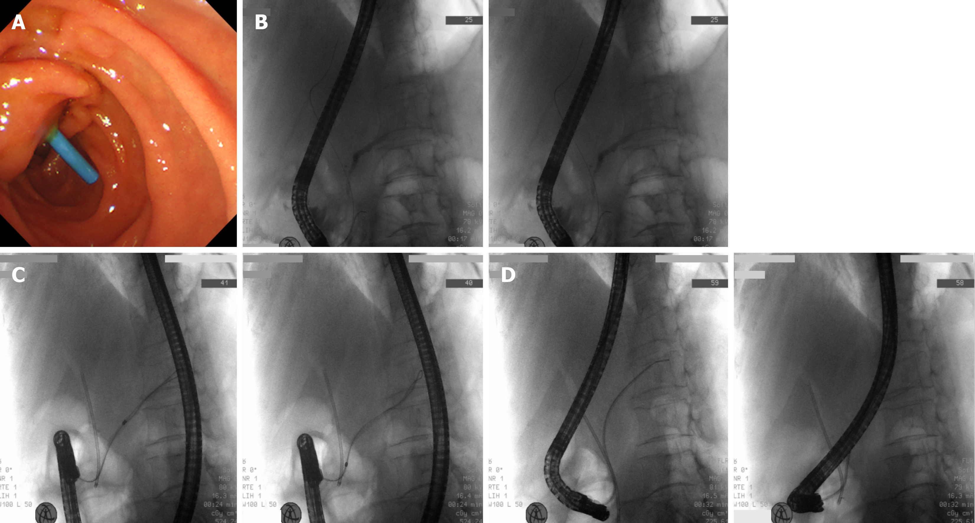
Figure 2 Sequential deployment of pancreatic stent under combined endoscopic and fluoroscopic surveillance in Case 2.
A: Endoscopic view of completed minor papilla cannulation with the catheter positioned inside the orifice for pancreatic stent delivery; B: Initial fluoroscopic localization: Guidewire anchored deep within the pancreatic tail to provide stable rail for stent advancement; C: Fluoroscopic monitoring during stent propulsion: The single-pigtail pancreatic stent is advanced along the guidewire with coordinated nursing instrument preparation; D: Final fluoroscopic confirmation after full stent release; dual endoscopic-fluoroscopic inspection confirms appropriate stent location and satisfactory pancreatic drainage.
- Citation: Jin HB, Huang JH, Wang SS, Dai SX, Li Y, Li LF. Nursing management for patients with minor papilla stent placement after failed endoscopic retrograde cholangiopancreatography cannulation: Two case reports. World J Gastrointest Endosc 2026; 18(11): 126966
- URL: https://www.wjgnet.com/1948-5190/full/v18/i11/126966.htm
- DOI: https://dx.doi.org/10.4253/wjge.126966