
















©The Author(s) 2025.
World J Gastrointest Endosc. Sep 16, 2025; 17(9): 110850
Published online Sep 16, 2025. doi: 10.4253/wjge.v17.i9.110850
Published online Sep 16, 2025. doi: 10.4253/wjge.v17.i9.110850
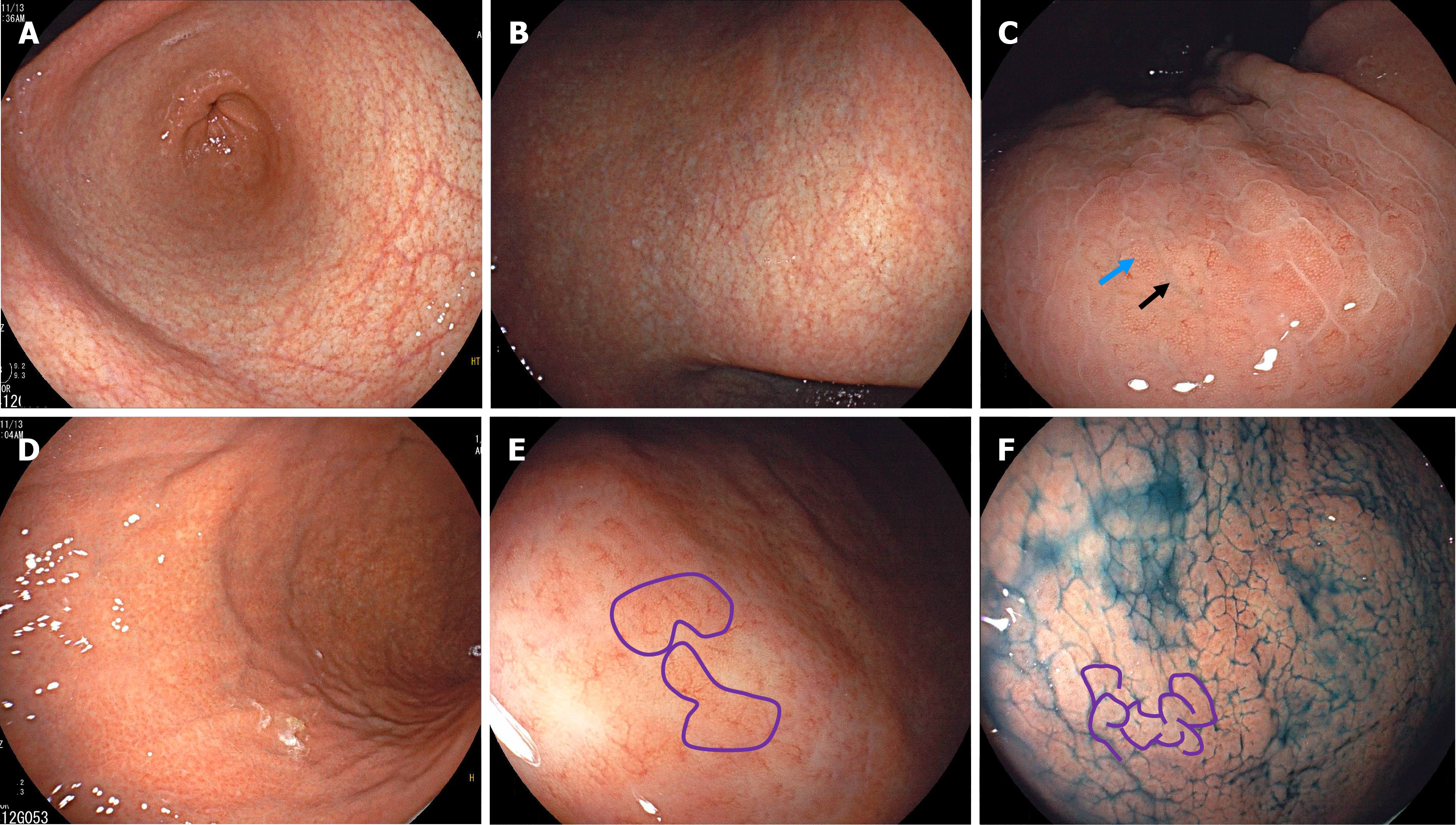
Figure 3 Endoscopic images from the patient in Case 2.
A: Atrophic changes in the antrum; B: Atrophy extending toward the lesser curvature of the corpus; C: A gyrus-like pattern of mild mucosal swelling (blue arrow) and deep fissures (black arrow) can be observed on the anterior wall of the corpus. There were visible collecting venules on the surface of the elevated mucosa; D: Alternating distribution of remnant oxyntic mucosa and atrophic mucosa on the greater curvature of the corpus; E: A close-up view showing remnant oxyntic mucosa with gyrus-like morphology (purple lines) on the greater curvature of the corpus; F: Indigo carmine staining highlighted the widespread gyrus-like changes (purple lines) in the greater curvature of the corpus.
- Citation: Tan CC, Shangguan XL, Lei XM, Deng FF, Wu YP, Zhang GM. Gyrus-like endoscopic changes in autoimmune gastritis with past Helicobacter pylori infection: Two case reports and review of literature. World J Gastrointest Endosc 2025; 17(9): 110850
- URL: https://www.wjgnet.com/1948-5190/full/v17/i9/110850.htm
- DOI: https://dx.doi.org/10.4253/wjge.v17.i9.110850