
















©The Author(s) 2025.
World J Gastrointest Endosc. Sep 16, 2025; 17(9): 110850
Published online Sep 16, 2025. doi: 10.4253/wjge.v17.i9.110850
Published online Sep 16, 2025. doi: 10.4253/wjge.v17.i9.110850
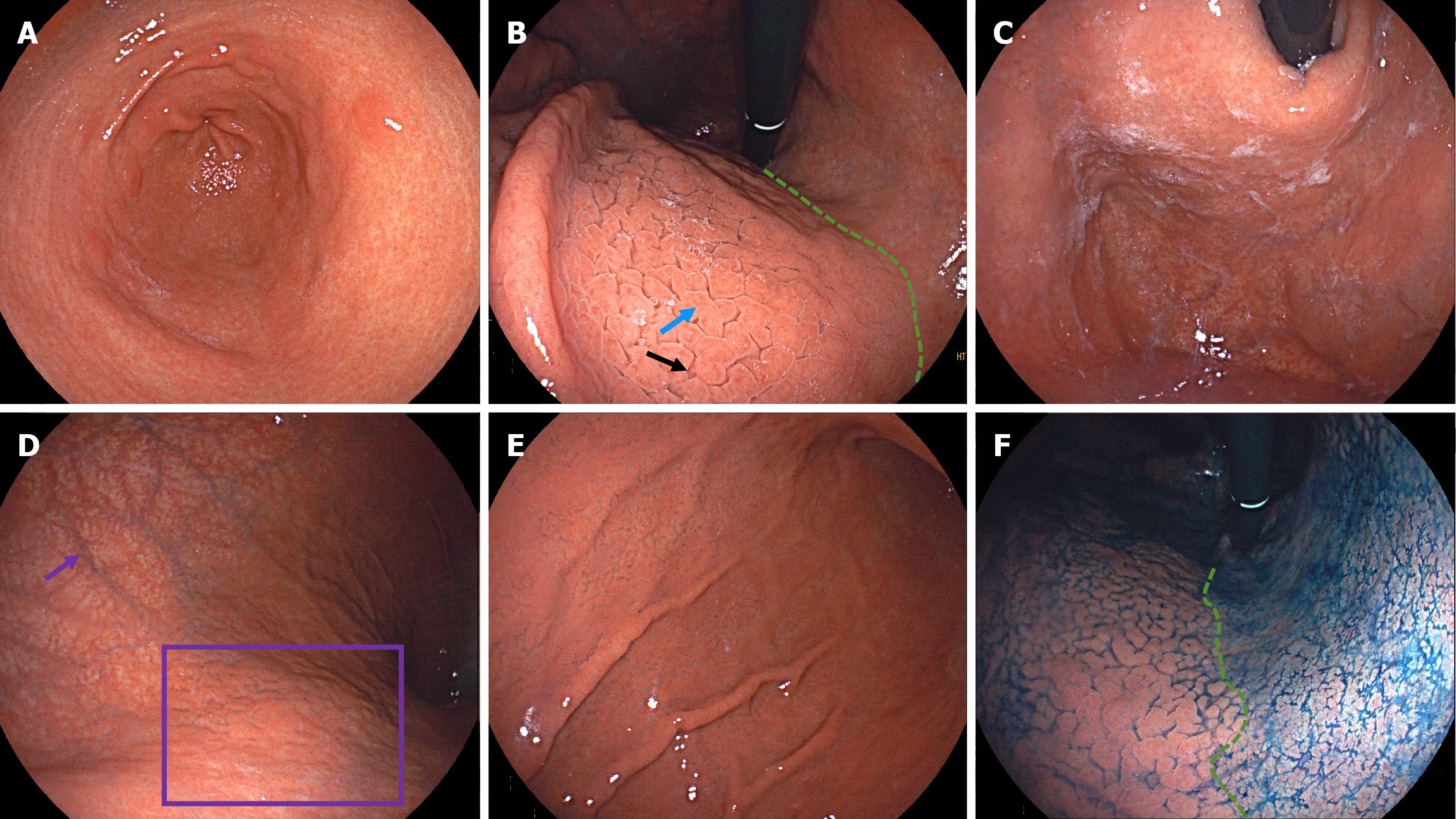
Figure 1 Endoscopic images from the patient in Case 1.
A: Atrophy of the antrum with visible xanthomas and circular wrinkle-like patterns; B: Atrophy along the lesser curvature of the corpus, with a clearly visible atrophic border (green line). Gyrus-like changes on the anterior wall of the corpus in the form of mucosal swelling (blue arrow) and deep fissures (black arrow); C: Adherent white mucus in the fundus; D: Severe atrophy in the upper greater curvature of the corpus, with visible submucosal vessels (purple arrow) after adequate insufflation. Scattered grid-like atrophy (purple box) can be seen on the posterior wall; E: Gyrus-like changes in the mid-to-lower greater curvature of the corpus; F: Indigo carmine staining clearly highlighted the atrophic border.
- Citation: Tan CC, Shangguan XL, Lei XM, Deng FF, Wu YP, Zhang GM. Gyrus-like endoscopic changes in autoimmune gastritis with past Helicobacter pylori infection: Two case reports and review of literature. World J Gastrointest Endosc 2025; 17(9): 110850
- URL: https://www.wjgnet.com/1948-5190/full/v17/i9/110850.htm
- DOI: https://dx.doi.org/10.4253/wjge.v17.i9.110850