
















©The Author(s) 2025.
World J Gastrointest Endosc. Sep 16, 2025; 17(9): 110424
Published online Sep 16, 2025. doi: 10.4253/wjge.v17.i9.110424
Published online Sep 16, 2025. doi: 10.4253/wjge.v17.i9.110424
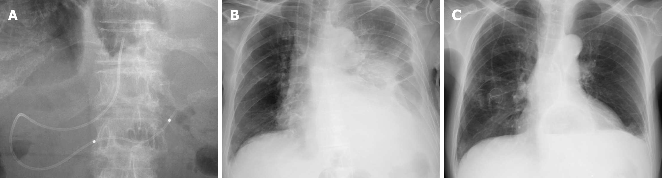
Figure 5 Internalization of the nasopancreatic drainage tube and changes in the pleural effusion volume.
A: An upper gastrointestinal endoscopy is performed to cut the endoscopic nasopancreatic drainage (ENPD) tube within the stomach using a Loop Cutter (Olympus, Tokyo, Japan); B: Chest radiograph at the time of admission. Left pleural effusion is observed; C: Chest radiograph 26 days after ENPD placement. The left pleural effusion is nearly resolved.
- Citation: Makazu M, Koizumi K, Kubota J, Kimura K, Masuda S. Transpapillary drainage of pancreatic fluid leakage via a rigid trans-tumoral tract using a drill dilator: A case report. World J Gastrointest Endosc 2025; 17(9): 110424
- URL: https://www.wjgnet.com/1948-5190/full/v17/i9/110424.htm
- DOI: https://dx.doi.org/10.4253/wjge.v17.i9.110424