
















©The Author(s) 2025.
World J Gastrointest Endosc. Sep 16, 2025; 17(9): 110424
Published online Sep 16, 2025. doi: 10.4253/wjge.v17.i9.110424
Published online Sep 16, 2025. doi: 10.4253/wjge.v17.i9.110424
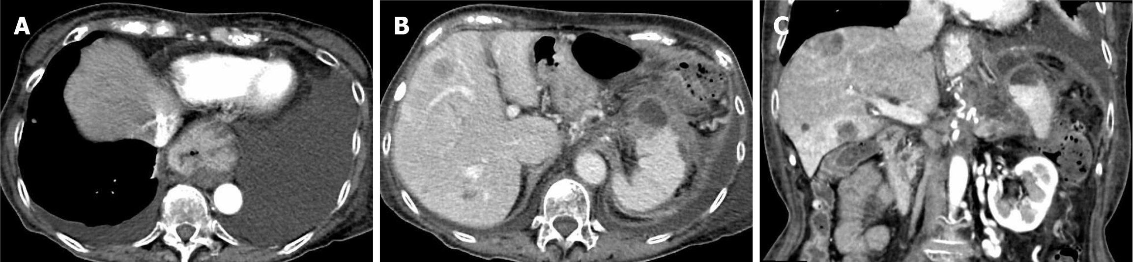
Figure 2 Dynamic computed tomography images of the pancreas at admission for pancreatic fluid leakage.
A: Axial chest view. A large amount of pleural effusion is observed in the left thoracic cavity, along with a small amount in the right thoracic cavity; B: Axial abdominal view; C: Coronal abdominal view. Perisplenic fluid accumulation with partial capsular-like structures is observed.
- Citation: Makazu M, Koizumi K, Kubota J, Kimura K, Masuda S. Transpapillary drainage of pancreatic fluid leakage via a rigid trans-tumoral tract using a drill dilator: A case report. World J Gastrointest Endosc 2025; 17(9): 110424
- URL: https://www.wjgnet.com/1948-5190/full/v17/i9/110424.htm
- DOI: https://dx.doi.org/10.4253/wjge.v17.i9.110424