
















©The Author(s) 2025.
World J Gastrointest Endosc. Sep 16, 2025; 17(9): 109144
Published online Sep 16, 2025. doi: 10.4253/wjge.v17.i9.109144
Published online Sep 16, 2025. doi: 10.4253/wjge.v17.i9.109144
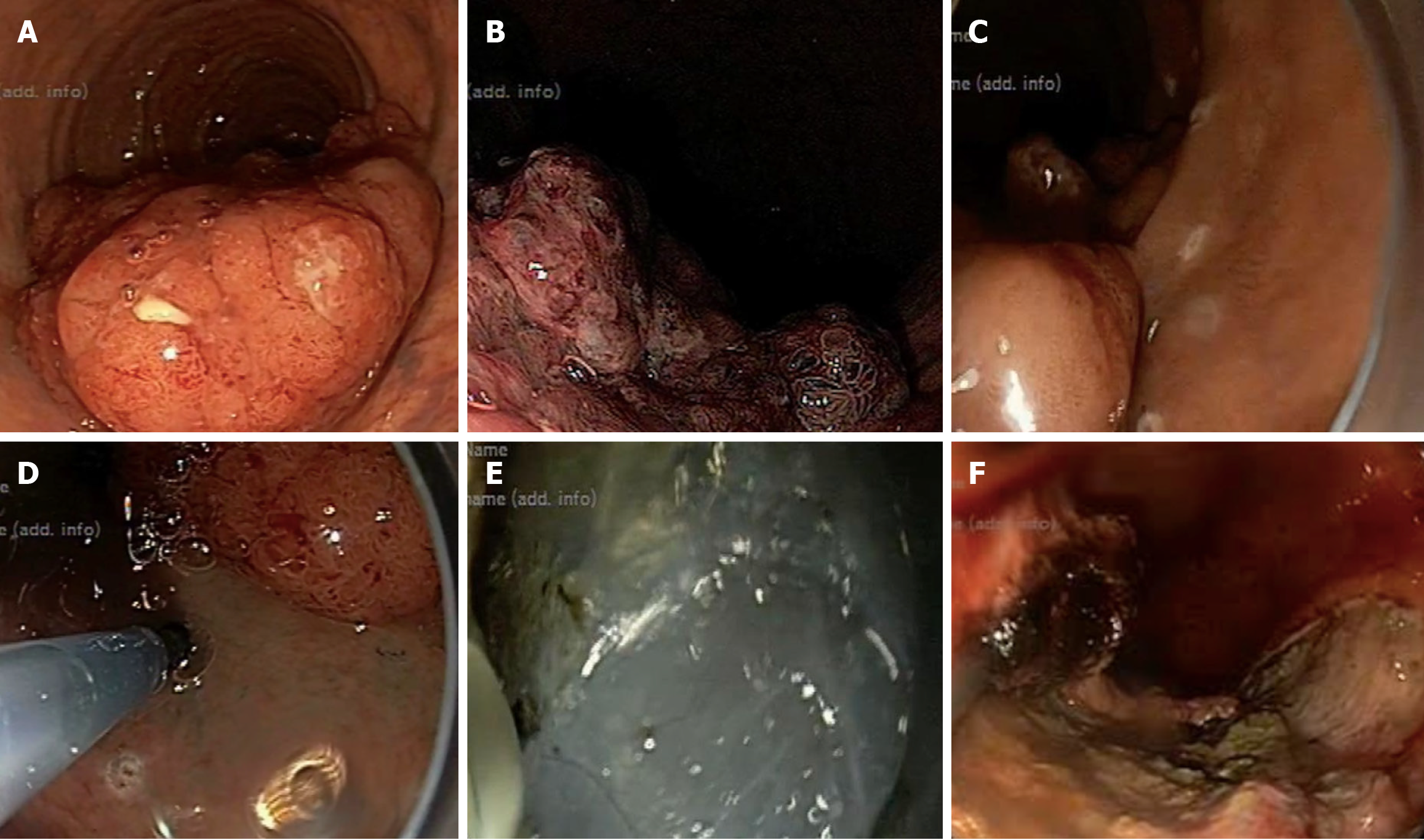
Figure 1 Steps of endoscopic submucosal dissection.
A: Endoscopic view of a rectal sessile polypoidal lesion; B: Narrow band imaging showing a Japanese Narrow Band Imaging Expert Team type 2B lesion; C: Marking around the lesion with a dual-knife; D: Submucosal injection with methylene blue; E: Submucosal dissection with a dual-knife; F: Base of the lesion after resection and one clip applied over a visible vessel to achieve hemostasis.
- Citation: Pal S, Bhaduri G. Endoscopic submucosal dissection for early gastrointestinal malignancies: Current state and future perspectives. World J Gastrointest Endosc 2025; 17(9): 109144
- URL: https://www.wjgnet.com/1948-5190/full/v17/i9/109144.htm
- DOI: https://dx.doi.org/10.4253/wjge.v17.i9.109144