
















©The Author(s) 2025.
World J Gastrointest Endosc. Sep 16, 2025; 17(9): 108549
Published online Sep 16, 2025. doi: 10.4253/wjge.v17.i9.108549
Published online Sep 16, 2025. doi: 10.4253/wjge.v17.i9.108549
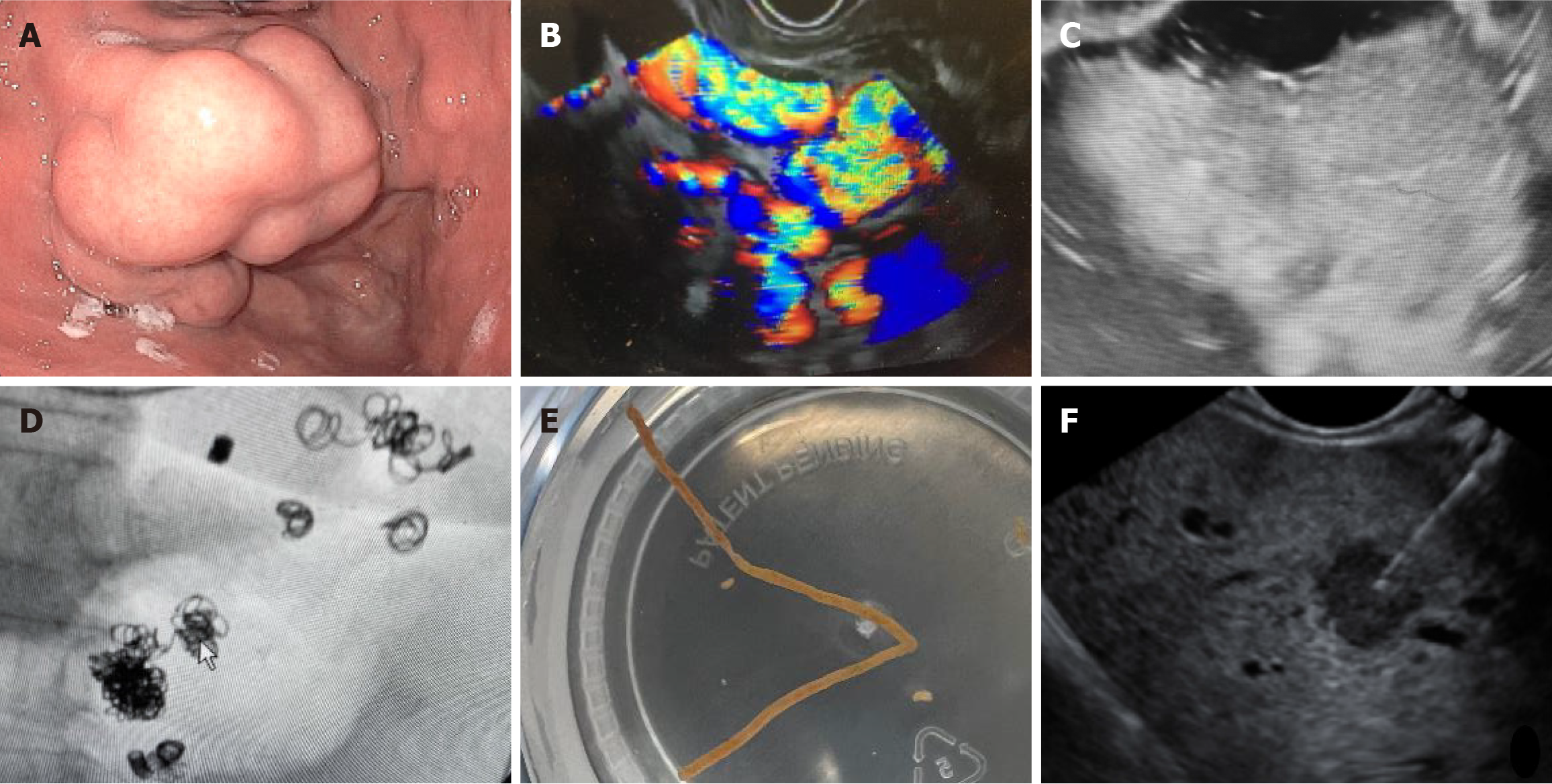
Figure 2 Endoscopic ultrasound appearance of different cirrhosis related conditions.
A: Endoscopic appearance of isolated gastric varix type 1 (IGV-1); B: Endoscopic ultrasound (EUS) appearance of isolated gastric varix type 1; C: EUS appearance of isolated gastric varix type 1 after glue injection (absence of doppler signal indicating obliteration); D: IGV after coil insertion (fluoroscopy); E: EUS guided liver biopsy sample; F: EUS guided biopsy of liver lesion.
- Citation: Castleberry DT, Mann R, Tharian B, Thandassery RB. Endoscopic ultrasound in the management of complications related to cirrhosis- recent evidence. World J Gastrointest Endosc 2025; 17(9): 108549
- URL: https://www.wjgnet.com/1948-5190/full/v17/i9/108549.htm
- DOI: https://dx.doi.org/10.4253/wjge.v17.i9.108549