
















©The Author(s) 2025.
World J Gastrointest Endosc. Sep 16, 2025; 17(9): 108420
Published online Sep 16, 2025. doi: 10.4253/wjge.v17.i9.108420
Published online Sep 16, 2025. doi: 10.4253/wjge.v17.i9.108420
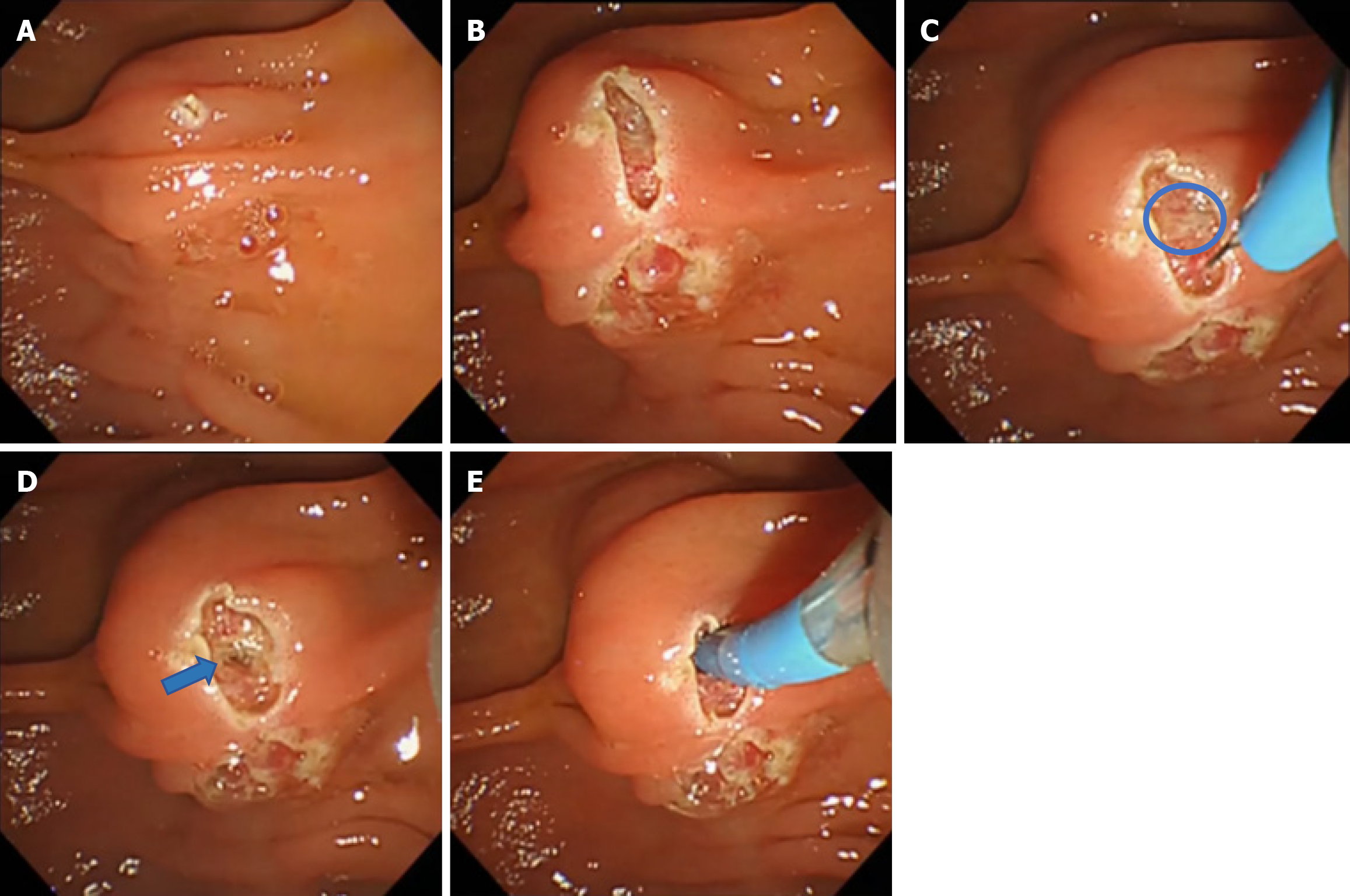
Figure 3 Stepwise endoscopic images of early precut needle knife fistulotomy in a case with a large oral protrusion.
A: Papilla with a long oral protrusion (oral protrusion-L); the incision line is marked at the apex of the protrusion prior to precutting; B: The initial incision is made using a needle knife, limited to the oral protrusion only, avoiding the papillary orifice characteristic of needle knife fistulotomy; C: Exposure of the sphincter of the Oddi muscle layer (highlighted with a blue circle). The extended oral protrusion allows for a wide incision plane, facilitating clear visualization of the muscle layer; D: Identification of the artificially created bile duct opening after further dissection (blue arrow); E: Successful bile duct cannulation with a guidewire through the exposed opening.
- Citation: Kaneko T, Kida M, Kurosu T, Kitahara G, Betto T, Saito Y, Koyama S, Nomura N, Kusano C. Early precut is useful for difficult bile duct cannulation, particularly in cases with long oral protrusion. World J Gastrointest Endosc 2025; 17(9): 108420
- URL: https://www.wjgnet.com/1948-5190/full/v17/i9/108420.htm
- DOI: https://dx.doi.org/10.4253/wjge.v17.i9.108420