Copyright
©The Author(s) 2025.
World J Gastrointest Endosc. Jul 16, 2025; 17(7): 107059
Published online Jul 16, 2025. doi: 10.4253/wjge.v17.i7.107059
Published online Jul 16, 2025. doi: 10.4253/wjge.v17.i7.107059
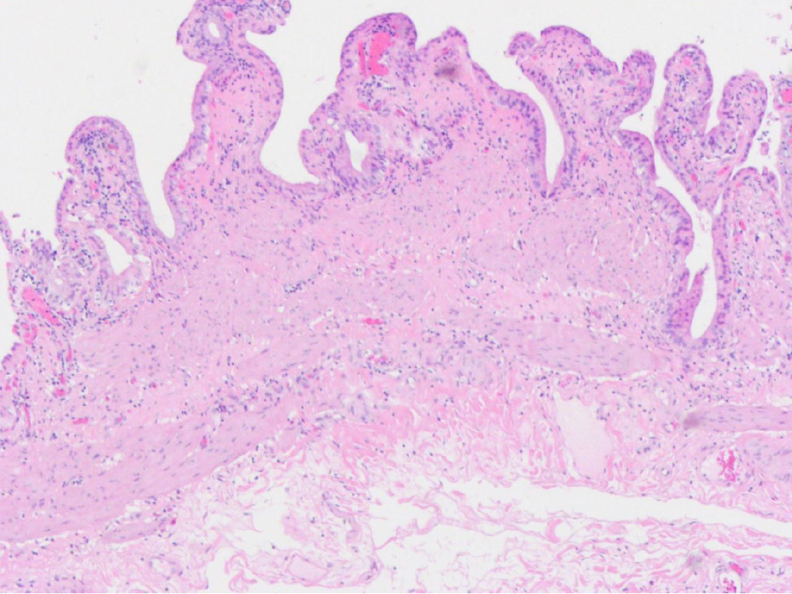
Figure 4 Pathology showed the thickness of the gallbladder wall was 0.
1-0.2 cm. The gallbladder was grayish-white, the mucosal surface was dark green, the mucosal surface was smooth, and the folds were visible. No significant abnormal pathological findings were found (hematoxylin-eosin staining, × 100).
- Citation: Wu JR, Wang CC, Li BY, Li JH, Zhang T, Li ZY. Concomitant functional gallbladder disorder and left-sided gallbladder: A case report. World J Gastrointest Endosc 2025; 17(7): 107059
- URL: https://www.wjgnet.com/1948-5190/full/v17/i7/107059.htm
- DOI: https://dx.doi.org/10.4253/wjge.v17.i7.107059


