Copyright
©The Author(s) 2025.
World J Gastrointest Endosc. Jul 16, 2025; 17(7): 105773
Published online Jul 16, 2025. doi: 10.4253/wjge.v17.i7.105773
Published online Jul 16, 2025. doi: 10.4253/wjge.v17.i7.105773
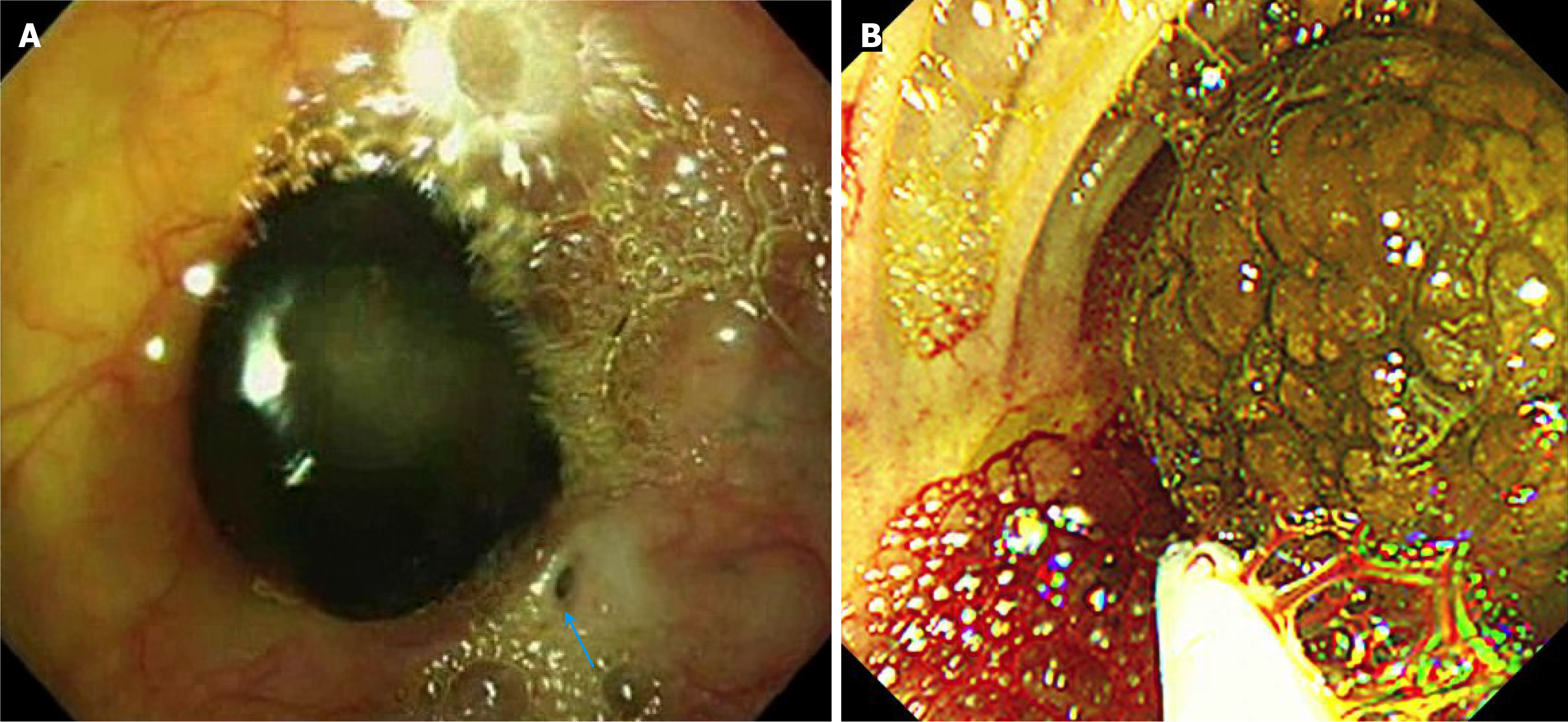
Figure 2 Intraoperative endoscopic view of stone.
A: Intraoperative endoscopic findings in an unpublished previous case of a Kasai portoenterostomy (KPE) site stone, experienced 14 years ago, showing a black pigmented stone hanging like a stalactite from the KPE site and a tiny opening of a bile ductule (arrow). Endoscopic removal of the stone resolved the cholangitis and prevented further development of cholangitis in this case; B: Intraoperative endoscopic findings in the current case showing a large thick bile sludge-like lump hanging from the KPE site, which was easily broken during endoscopic removal. Three minutes after starting the endoscope examination, a sudden fatal air embolism occurred, and the patient died.
- Citation: Shin SY, Yeon HJ, Lee SO, Lee JR, Leem G, Han SJ. Fatal air embolism during intestinal endoscopy in Kasai portoenterostomy for biliary atresia: A case report. World J Gastrointest Endosc 2025; 17(7): 105773
- URL: https://www.wjgnet.com/1948-5190/full/v17/i7/105773.htm
- DOI: https://dx.doi.org/10.4253/wjge.v17.i7.105773


