











BPG is committed to discovery and dissemination of knowledge
Case Report
©The Author(s) 2025.
World J Gastrointest Endosc. Dec 16, 2025; 17(12): 114836
Published online Dec 16, 2025. doi: 10.4253/wjge.v17.i12.114836
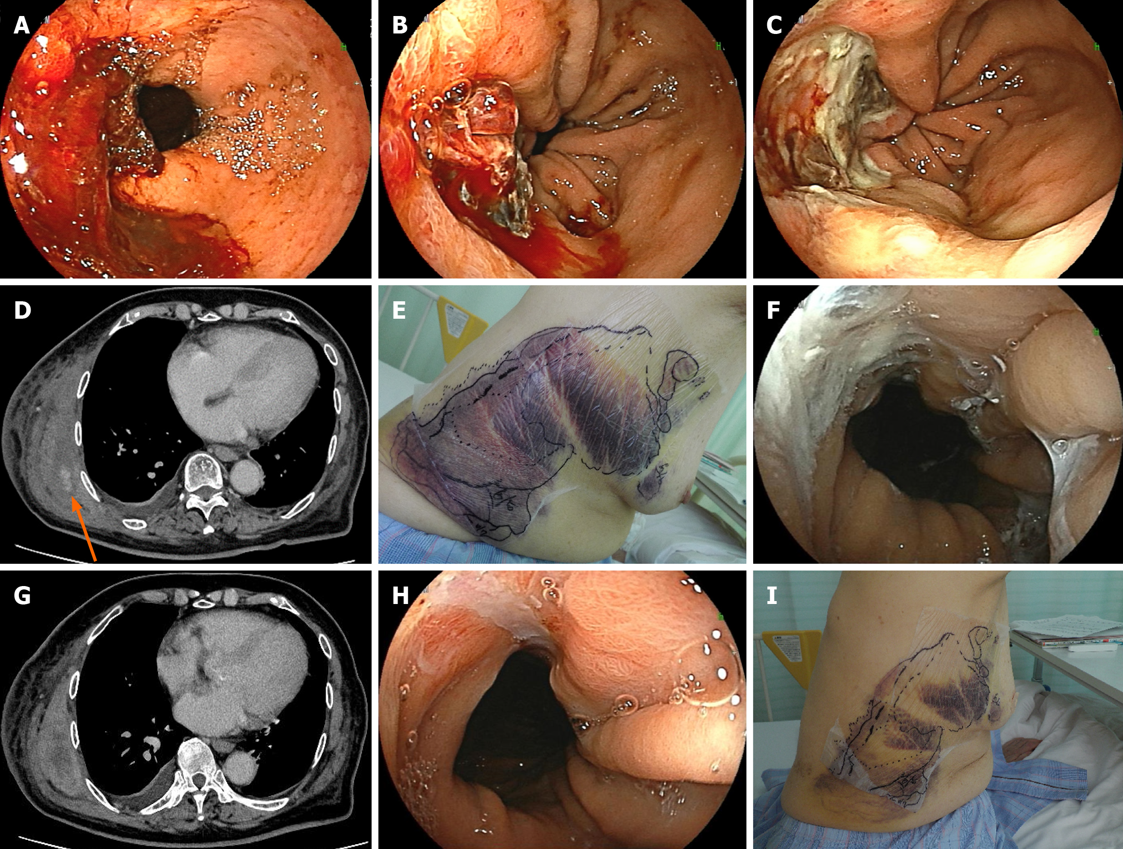
Figure 2
Figure 2 Management of refractory hemorrhage with a novel hemostatic agent: From diagnosis to hemostasis. A and B: Esophageal endoscopic findings at the time of admission. Before treatment, oozing hemorrhage was observed from an ulcer within the hiatal hernia (A); After treatment, clipping was attempted to stop the bleeding, but new bleeding occurred from the clipped area (B); C: Esophageal endoscopic findings of changes in ulcer lesions: A single dose of the hemostatic agent (Byclot®) resulted in complete hemostasis; D: Computed tomography findings of the intramuscular hematoma: A large intramuscular hematoma was present in the right chest, accompanied by arterial extravasation (indicated by the orange arrow); E: Subcutaneous bleeding finding: Widespread subcutaneous bleeding was observed in the right chest; F: Esophageal endoscopic findings of changes in ulcer lesions: Two weeks after the administration of Byclot® (around the same time as D and E), the esophageal ulcer remained hemostatic; G: Computed tomography findings of the intramuscular hematoma: Hemostasis was confirmed 5 days after the administration of a mixture of factors VIIa and X (Byclot®); H: Esophageal endoscopic findings of changes in ulcer lesions: One week after emicizumab administration, the esophageal ulcer showed a marked tendency to heal; I: Subcutaneous bleeding finding: One week after emicizumab administration, the subcutaneous bleeding resolved rapidly.


Write to the Help Desk