
















©The Author(s) 2025.
World J Gastrointest Endosc. Dec 16, 2025; 17(12): 111614
Published online Dec 16, 2025. doi: 10.4253/wjge.v17.i12.111614
Published online Dec 16, 2025. doi: 10.4253/wjge.v17.i12.111614
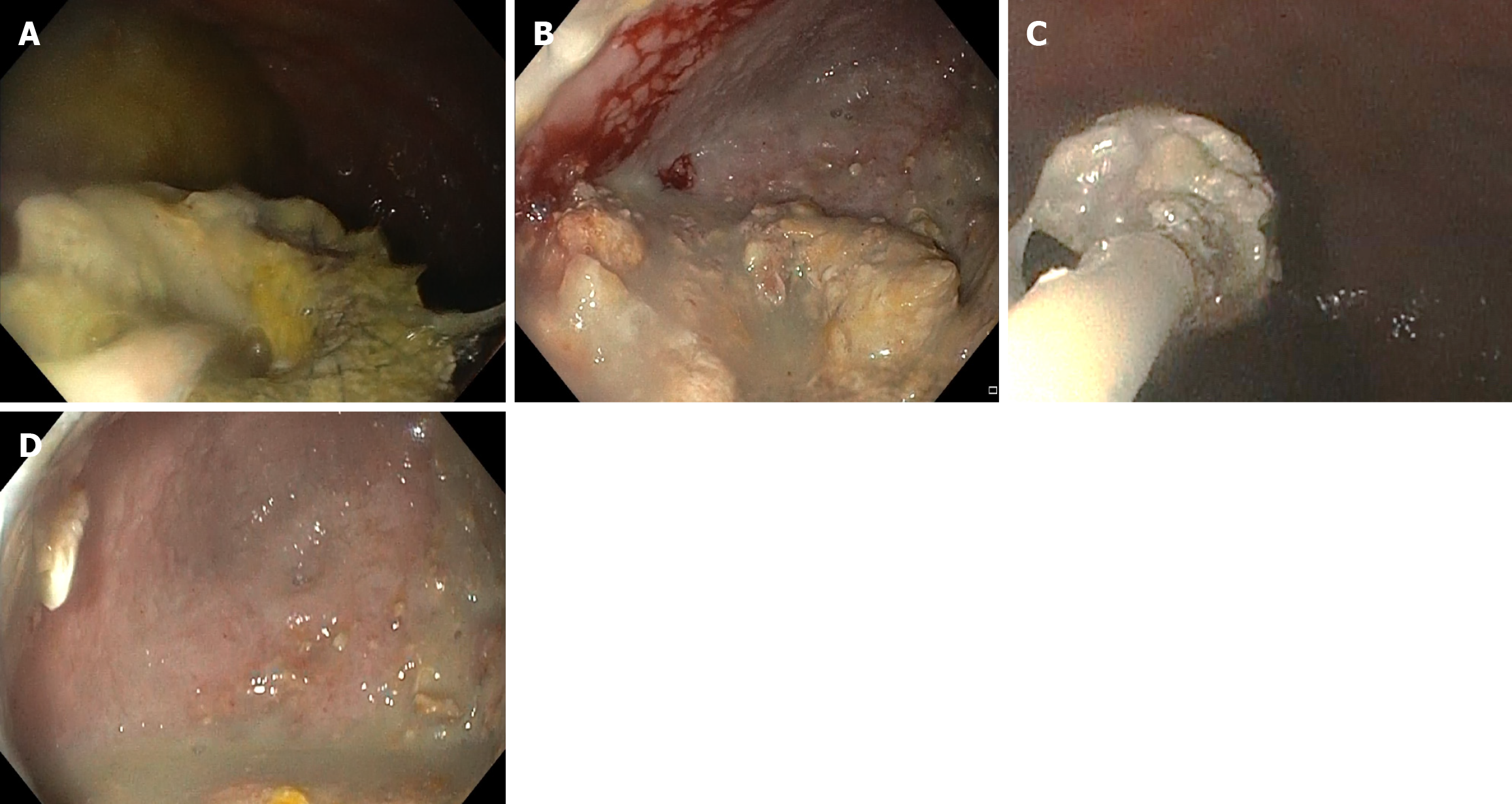
Figure 2 The procedure of direct endoscopic necrosectomy.
A: Endoscopic image shows blocked lumen-apposing metallic stents with pus seen oozing through it; B: Blockage in the lumen-apposing metallic stents is removed, and the scope is negotiated into the cavity, which shows a large cavity with necrotic debris; C: Necrotic debris is removed with Roth net forceps; D: Post-necrosectomy with minimal debris and healthy wall cavity seen.
- Citation: Lone SA, Sonika U, Reddy RT, Vaithiyam V, Aneesh PS, Palli SH, Dalal A, Kumar A, Srivastava S, Sachdeva S. Predictive factors and outcomes of endoscopic necrosectomy in patients with acute pancreatitis and walled-off necrosis. World J Gastrointest Endosc 2025; 17(12): 111614
- URL: https://www.wjgnet.com/1948-5190/full/v17/i12/111614.htm
- DOI: https://dx.doi.org/10.4253/wjge.v17.i12.111614