
















©The Author(s) 2025.
World J Gastrointest Endosc. Dec 16, 2025; 17(12): 111614
Published online Dec 16, 2025. doi: 10.4253/wjge.v17.i12.111614
Published online Dec 16, 2025. doi: 10.4253/wjge.v17.i12.111614
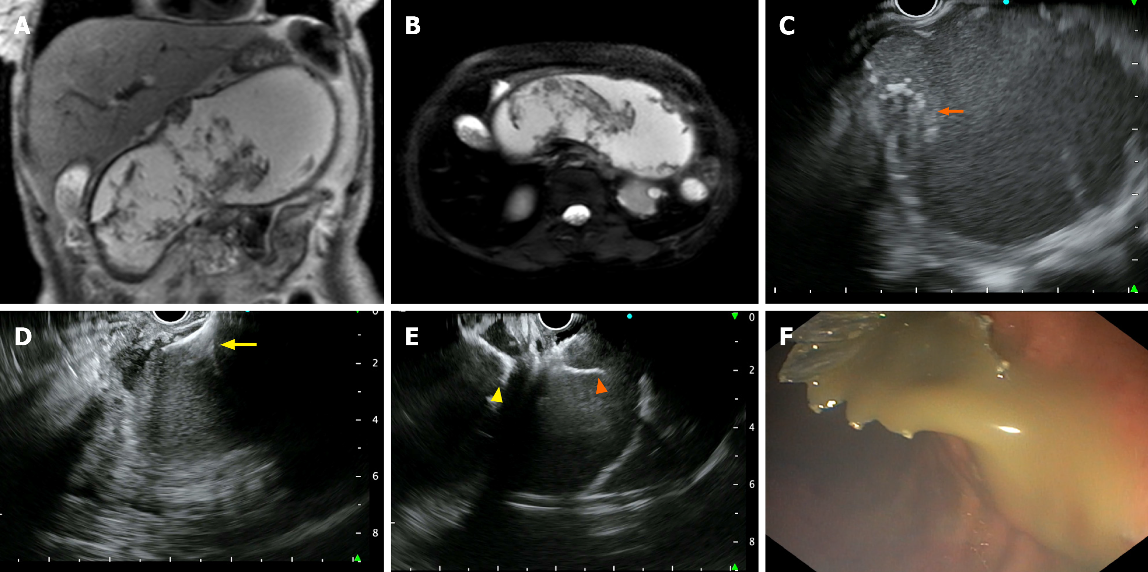
Figure 1 Magnetic resonance imaging and endoscopic ultrasound guided drainage of lumen-apposing metallic stents.
A: Magnetic resonance T2-weighted imaging coronal; B: Axial views showing walled-off necrosis of the pancreas with significant debris; C: Endoscopic ultrasound (EUS) reveals a large walled-off necrosis with echogenic contents and debris (orange arrow); D: EUS-guided lumen-apposing metallic stents (LAMS) are being placed, with distal flanges (yellow arrow) deployed; E: Both distal flanges (yellow arrowhead) inside the cavity and proximal flanges (orange arrowhead) in the gastric cavity are deployed seen in EUS; F: Endoscopic image post-deployment of LAMS and pus seen oozing out from out the LAMS.
- Citation: Lone SA, Sonika U, Reddy RT, Vaithiyam V, Aneesh PS, Palli SH, Dalal A, Kumar A, Srivastava S, Sachdeva S. Predictive factors and outcomes of endoscopic necrosectomy in patients with acute pancreatitis and walled-off necrosis. World J Gastrointest Endosc 2025; 17(12): 111614
- URL: https://www.wjgnet.com/1948-5190/full/v17/i12/111614.htm
- DOI: https://dx.doi.org/10.4253/wjge.v17.i12.111614