
















©The Author(s) 2025.
World J Gastrointest Endosc. Dec 16, 2025; 17(12): 110594
Published online Dec 16, 2025. doi: 10.4253/wjge.v17.i12.110594
Published online Dec 16, 2025. doi: 10.4253/wjge.v17.i12.110594
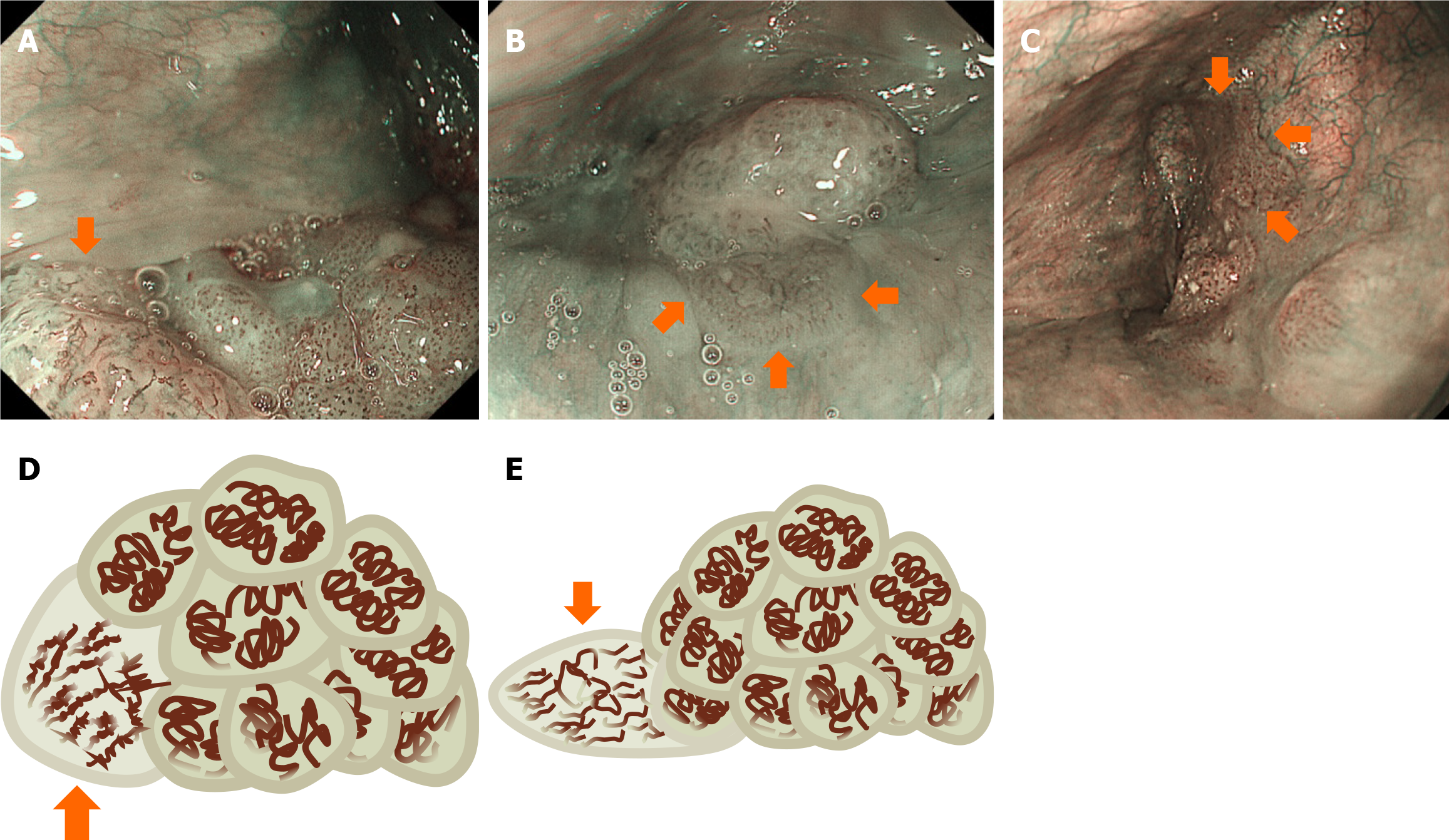
Figure 3 Typical endoscopic images of oral and pharyngolaryngeal squamous cell carcinomas with elevated morphology.
A-C: Typical endoscopic images; D and E: The schema. A and D: In a squamous cell carcinoma lesion, an uneven, lobulated structure and irregular morphology of capillaries are observed (arrow); B and E: Coexistence of a flat lesion is evident within a squamous cell carcinoma (arrows). The lobulated structure is absent, and the morphology of the capillaries is irregular in the flat lesion; C: Another squamous cell carcinoma accompanies a flat lesion without a lobulated structure (arrows).
- Citation: Iwamuro M, Tanaka T, Hamada K, Kono Y, Kawano S, Kawahara Y, Otsuka M. Endoscopic features of oral and pharyngolaryngeal papillomas and their role in distinguishing squamous cell carcinoma. World J Gastrointest Endosc 2025; 17(12): 110594
- URL: https://www.wjgnet.com/1948-5190/full/v17/i12/110594.htm
- DOI: https://dx.doi.org/10.4253/wjge.v17.i12.110594