
















©The Author(s) 2025.
World J Gastrointest Endosc. Dec 16, 2025; 17(12): 110594
Published online Dec 16, 2025. doi: 10.4253/wjge.v17.i12.110594
Published online Dec 16, 2025. doi: 10.4253/wjge.v17.i12.110594
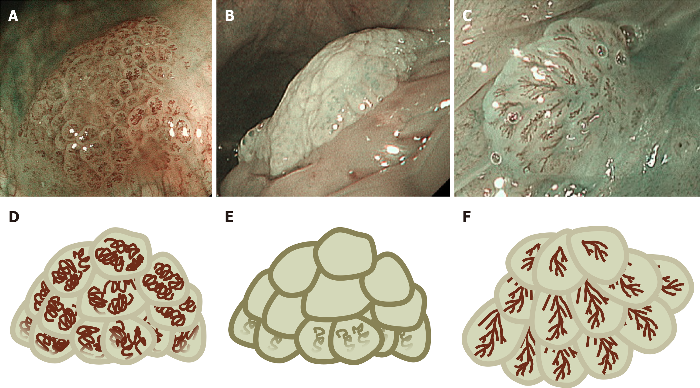
Figure 1 Classification of oral and pharyngolaryngeal papillomas on narrow band and blue laser imaging.
A-C: Typical endoscopic images of oral and pharyngolaryngeal papillomas; D-F: Their corresponding schema. We classified oral and pharyngolaryngeal papillomas into three distinct patterns based on endoscopic features. A and D: Salmon roe-like polyp is an elevated lesion with aggregation of multiple small nodules. Coil-shaped capillaries are internally visible in each nodule, resembling salmon roe or frog eggs; B and E: A polyp without capillary transparency is similar to the previous type of polyp and involves an elevated lesion with aggregation of multiple small nodules. However, there is no capillary transparency or only slightly visible capillaries; C and F: Pinecone-like polyp resembles a pinecone. This type of polyp consists of overlapping scale-like components, and capillaries resembling leaf veins are visible in each element.
- Citation: Iwamuro M, Tanaka T, Hamada K, Kono Y, Kawano S, Kawahara Y, Otsuka M. Endoscopic features of oral and pharyngolaryngeal papillomas and their role in distinguishing squamous cell carcinoma. World J Gastrointest Endosc 2025; 17(12): 110594
- URL: https://www.wjgnet.com/1948-5190/full/v17/i12/110594.htm
- DOI: https://dx.doi.org/10.4253/wjge.v17.i12.110594