
















©The Author(s) 2025.
World J Gastrointest Endosc. Dec 16, 2025; 17(12): 110168
Published online Dec 16, 2025. doi: 10.4253/wjge.v17.i12.110168
Published online Dec 16, 2025. doi: 10.4253/wjge.v17.i12.110168
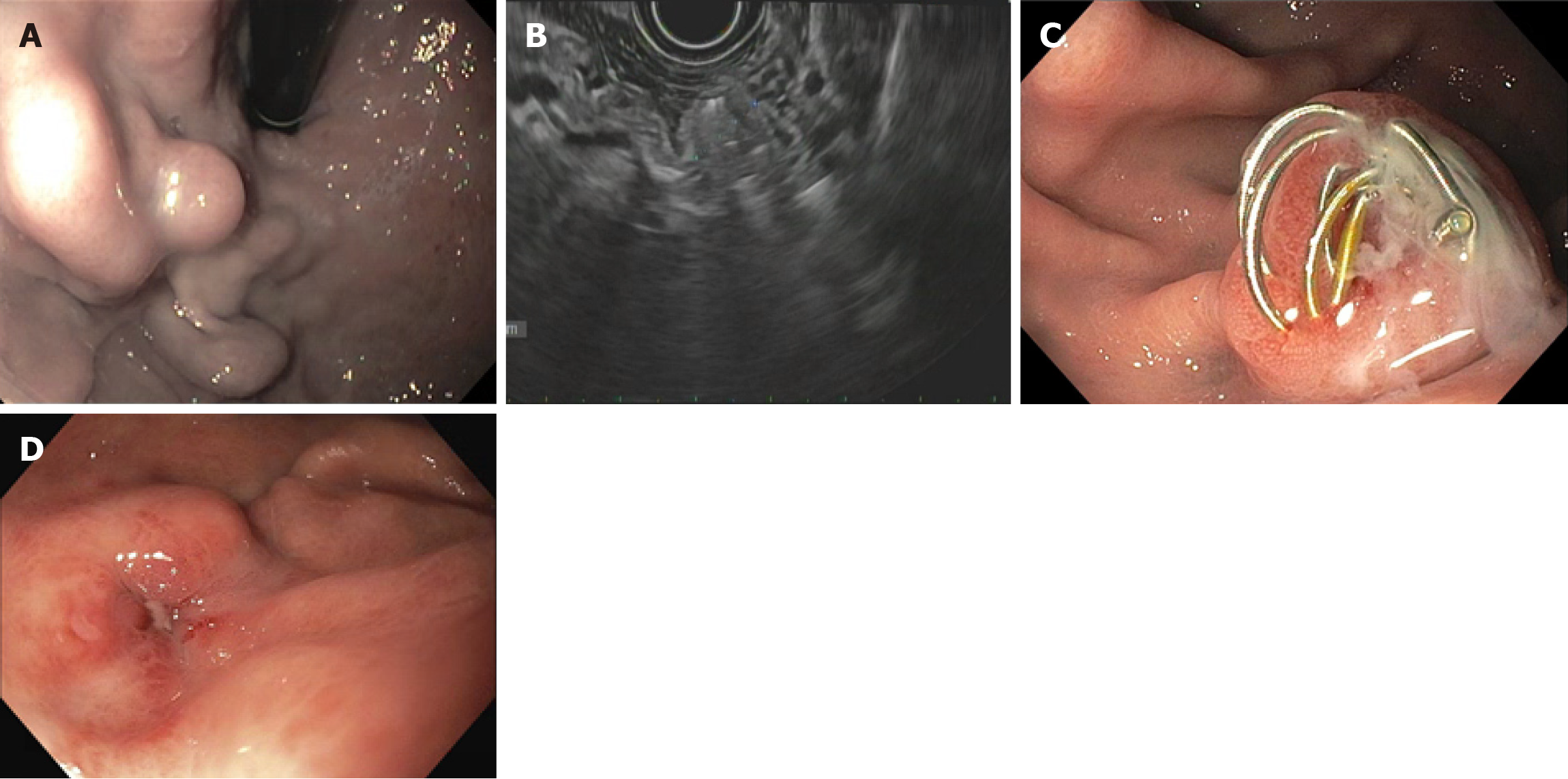
Figure 3 24-year-old male with history of portal vein thrombosis and hepatic encephalopathy was recently hospitalized with hematemesis and found to have gastric varices on upper endoscopy.
Imaging showed no evidence of gastro-renal shunt. He was referred for endoscopic ultrasound (EUS) guided therapy of gastric varices. A: Upper endoscopy showing isolated gastric varices type 1; B: EUS showing complete thrombosis of varices following EUS guided coil embolization and cyanoacrylate glue injection; C: Coil extrusion on follow-up endoscopy at 3 months; D: Follow-up endoscopy at 1 year showing complete eradication of varices.
- Citation: El Dada A, El Khoury M, Stephan P, Nehme F. Endoscopic ultrasound-guided coil embolization for gastric varices: A promising alternative to traditional therapies. World J Gastrointest Endosc 2025; 17(12): 110168
- URL: https://www.wjgnet.com/1948-5190/full/v17/i12/110168.htm
- DOI: https://dx.doi.org/10.4253/wjge.v17.i12.110168