
















©The Author(s) 2025.
World J Gastrointest Endosc. Dec 16, 2025; 17(12): 110168
Published online Dec 16, 2025. doi: 10.4253/wjge.v17.i12.110168
Published online Dec 16, 2025. doi: 10.4253/wjge.v17.i12.110168
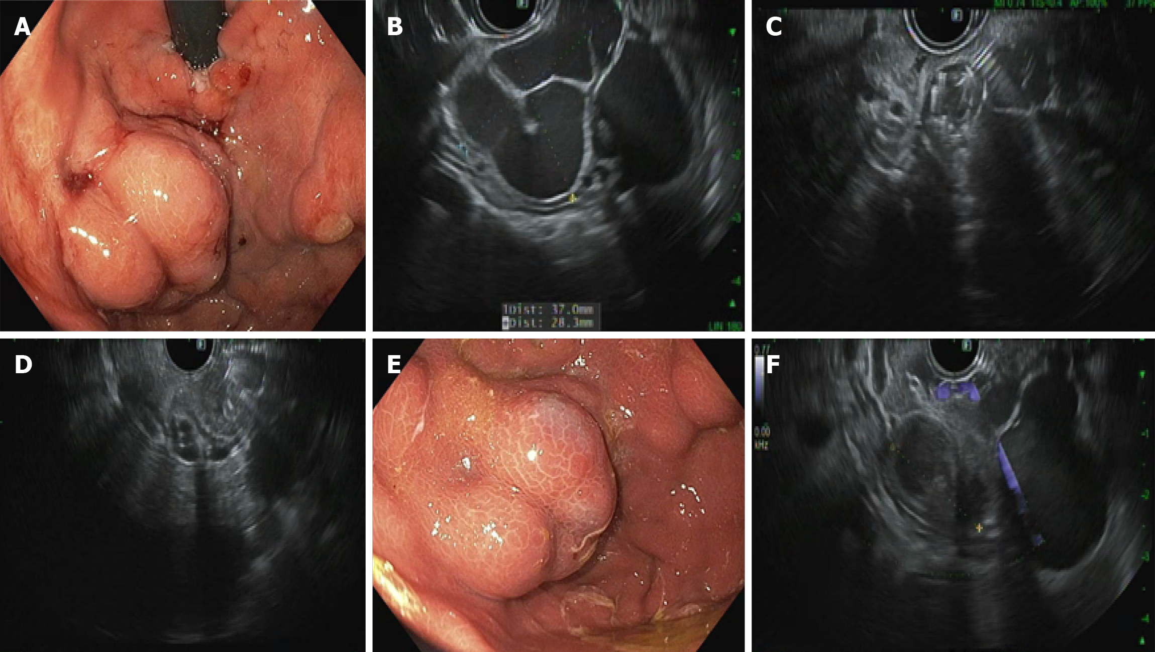
Figure 1 32-year-old male with history of cirrhosis secondary to alcohol use was referred for primary prophylaxis of gastric variceal bl
- Citation: El Dada A, El Khoury M, Stephan P, Nehme F. Endoscopic ultrasound-guided coil embolization for gastric varices: A promising alternative to traditional therapies. World J Gastrointest Endosc 2025; 17(12): 110168
- URL: https://www.wjgnet.com/1948-5190/full/v17/i12/110168.htm
- DOI: https://dx.doi.org/10.4253/wjge.v17.i12.110168