
















©The Author(s) 2025.
World J Gastrointest Endosc. Oct 16, 2025; 17(10): 111408
Published online Oct 16, 2025. doi: 10.4253/wjge.v17.i10.111408
Published online Oct 16, 2025. doi: 10.4253/wjge.v17.i10.111408
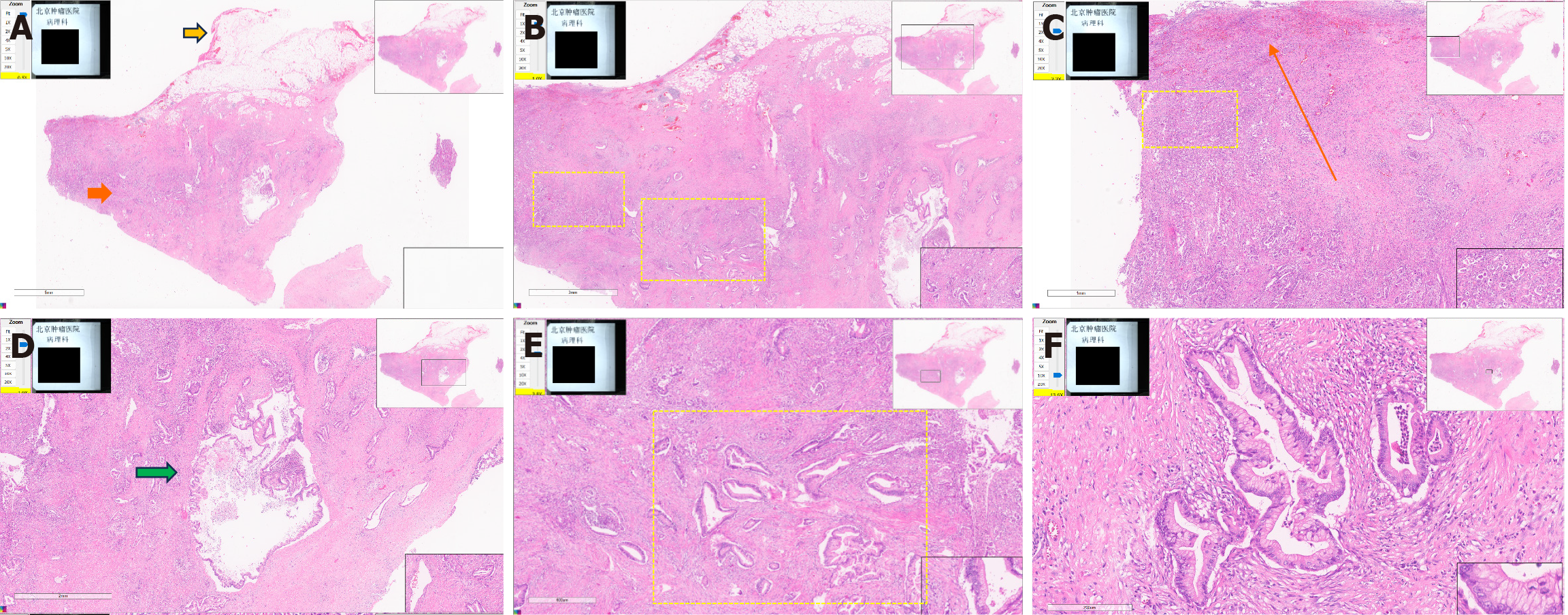
Figure 5 Histopathological analysis of a resected specimen from a 58-year-old woman with a gastric mass.
A-F: Histopathology confirmed a nonpancreatic primary adenocarcinoma with predominant gastric involvement. The lesion exhibited cystic degeneration and extended to the pyloroduodenal region, forming mucosal ulcers. Representative section demonstrating the lesion’s architecture, including the serosal layer (yellow arrow) and the muscularis propria (orange arrow) (A). Infiltrative irregular glands (yellow dashed box) exhibiting malignant cytological features (nuclear pleomorphism and a crowded growth pattern) were observed within the muscularis propria (B and E). Tumor invasion through the mucosal to subserosal layers (orange arrow) of the gastrointestinal wall was apparent, with multiple tumorous cells and glands in the muscularis propria (yellow dashed box) (C). Cystic dilation (green arrow) of the ectopic pancreatic ductal structures (D). A high-power view revealed glandular epithelial cells with abundant eosinophilic cytoplasm and elongated nuclei, consistent with pancreatobiliary adenocarcinoma (F).
- Citation: Wang YX, Wang J, Liang SX, Wu Q. Primary adenocarcinoma from a gastric heterotopic pancreas: A case report. World J Gastrointest Endosc 2025; 17(10): 111408
- URL: https://www.wjgnet.com/1948-5190/full/v17/i10/111408.htm
- DOI: https://dx.doi.org/10.4253/wjge.v17.i10.111408