
















©The Author(s) 2025.
World J Gastrointest Endosc. Oct 16, 2025; 17(10): 111408
Published online Oct 16, 2025. doi: 10.4253/wjge.v17.i10.111408
Published online Oct 16, 2025. doi: 10.4253/wjge.v17.i10.111408
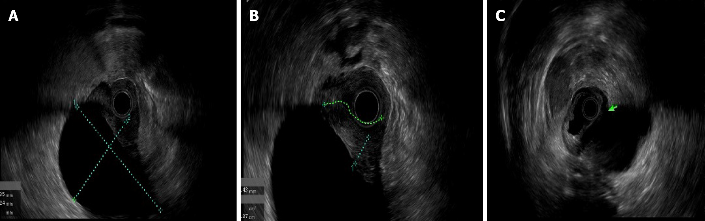
Figure 4 Endosonographic features of an abdominal solid cystic mass in a 58-year-old woman.
A: A multilobulated solid cystic mass (7.0 cm × 4.8 cm) was identified; B: The mass extended to the duodenal bulb-descending junction, causing luminal stenosis. The duodenal wall showed tumor infiltration (irregular hypoechoic thickening of up to 1 cm). The cyst wall exhibited hypoechoic unevenness, suggesting mural involvement; C: The lesion had ill-defined borders with the fourth layer of the gastric wall (as indicated by green arrow).
- Citation: Wang YX, Wang J, Liang SX, Wu Q. Primary adenocarcinoma from a gastric heterotopic pancreas: A case report. World J Gastrointest Endosc 2025; 17(10): 111408
- URL: https://www.wjgnet.com/1948-5190/full/v17/i10/111408.htm
- DOI: https://dx.doi.org/10.4253/wjge.v17.i10.111408