
















©The Author(s) 2025.
World J Gastrointest Endosc. Oct 16, 2025; 17(10): 111408
Published online Oct 16, 2025. doi: 10.4253/wjge.v17.i10.111408
Published online Oct 16, 2025. doi: 10.4253/wjge.v17.i10.111408
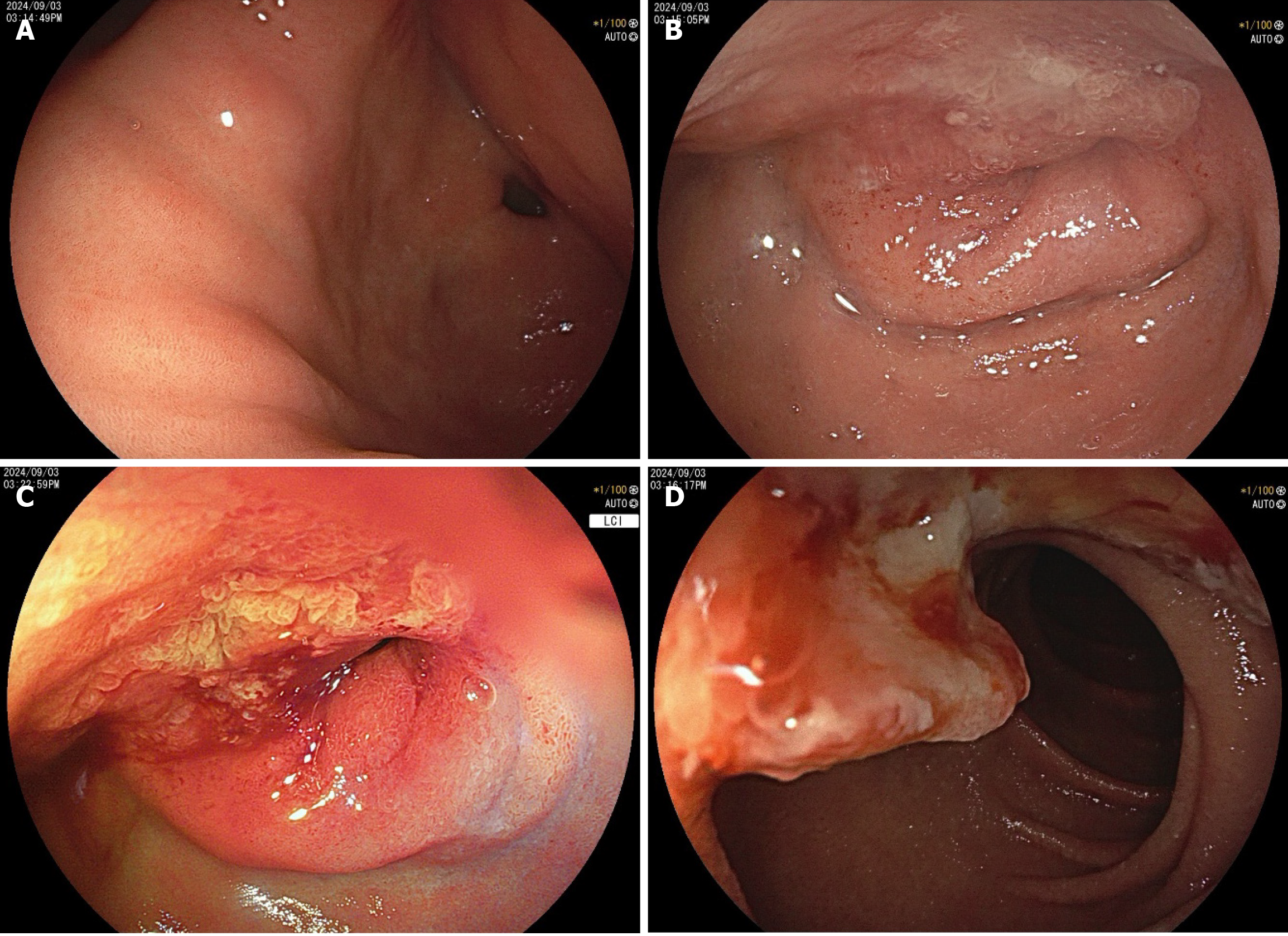
Figure 3 Endoscopic findings of ectopic pancreatic carcinoma in a 58-year-old woman.
A: A submucosal lesion with central umbilication was observed at the greater curvature of the gastric antrum, accompanied by extrinsic compression near the pylorus (soft on probing, with pyloric patency preserved); B and C: A giant ulcer with raised margins and a white fibrinoid base was found at the duodenal bulb-descending junction, causing significant luminal stenosis; D: The stenosis was nearly obstructive, the tumor in the descending duodenum exhibited mucosal breach and a coarse, irregular surface.
- Citation: Wang YX, Wang J, Liang SX, Wu Q. Primary adenocarcinoma from a gastric heterotopic pancreas: A case report. World J Gastrointest Endosc 2025; 17(10): 111408
- URL: https://www.wjgnet.com/1948-5190/full/v17/i10/111408.htm
- DOI: https://dx.doi.org/10.4253/wjge.v17.i10.111408