
















©The Author(s) 2025.
World J Gastrointest Endosc. Oct 16, 2025; 17(10): 111408
Published online Oct 16, 2025. doi: 10.4253/wjge.v17.i10.111408
Published online Oct 16, 2025. doi: 10.4253/wjge.v17.i10.111408
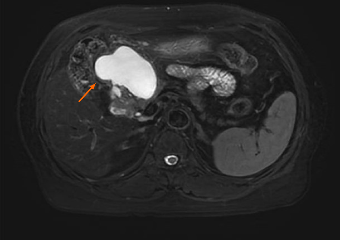
Figure 2 Contrast-enhanced magnetic resonance imaging findings in a 58-year-old woman with elevated carbohydrate antigen 19-9.
Magnetic resonance imaging revealed a well-circumscribed, oval cystic lesion (62 mm × 38 mm, SE3 IM9) in the right upper abdomen, with prolonged T2 signal intensity. The lesion was closely adjacent to the pancreatic head and the gastric antrum, with no fat plane separation. Peripheral wall enhancement was observed postcontrast.
- Citation: Wang YX, Wang J, Liang SX, Wu Q. Primary adenocarcinoma from a gastric heterotopic pancreas: A case report. World J Gastrointest Endosc 2025; 17(10): 111408
- URL: https://www.wjgnet.com/1948-5190/full/v17/i10/111408.htm
- DOI: https://dx.doi.org/10.4253/wjge.v17.i10.111408