
















©The Author(s) 2025.
World J Gastrointest Endosc. Oct 16, 2025; 17(10): 111408
Published online Oct 16, 2025. doi: 10.4253/wjge.v17.i10.111408
Published online Oct 16, 2025. doi: 10.4253/wjge.v17.i10.111408
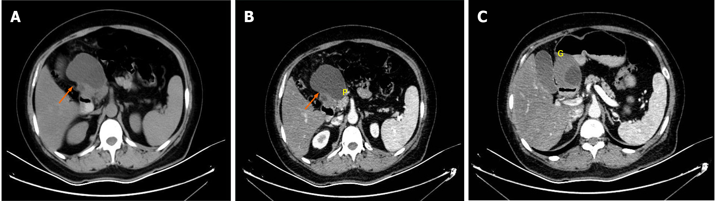
Figure 1 Abdominal computed tomography findings in a 58-year-old woman with elevated carbohydrate antigen 19-9.
A: The non-contrast-enhanced coronal view demonstrated a 7.1 cm × 4.8 cm hypodense cystic lesion (orange arrow) in the right upper quadrant; B and C: An arterial-phase contrast-enhanced coronal image reveals the lesion’s (orange arrow) intimate anatomical relationship with both the pancreatic head (P in B) and the greater curvature of the gastric antrum (G in C). The absence of a septum was found, and no significant arterial enhancement or suspicious nodularity of the cyst wall was found.
- Citation: Wang YX, Wang J, Liang SX, Wu Q. Primary adenocarcinoma from a gastric heterotopic pancreas: A case report. World J Gastrointest Endosc 2025; 17(10): 111408
- URL: https://www.wjgnet.com/1948-5190/full/v17/i10/111408.htm
- DOI: https://dx.doi.org/10.4253/wjge.v17.i10.111408