
















©The Author(s) 2025.
World J Gastrointest Endosc. Oct 16, 2025; 17(10): 110417
Published online Oct 16, 2025. doi: 10.4253/wjge.v17.i10.110417
Published online Oct 16, 2025. doi: 10.4253/wjge.v17.i10.110417
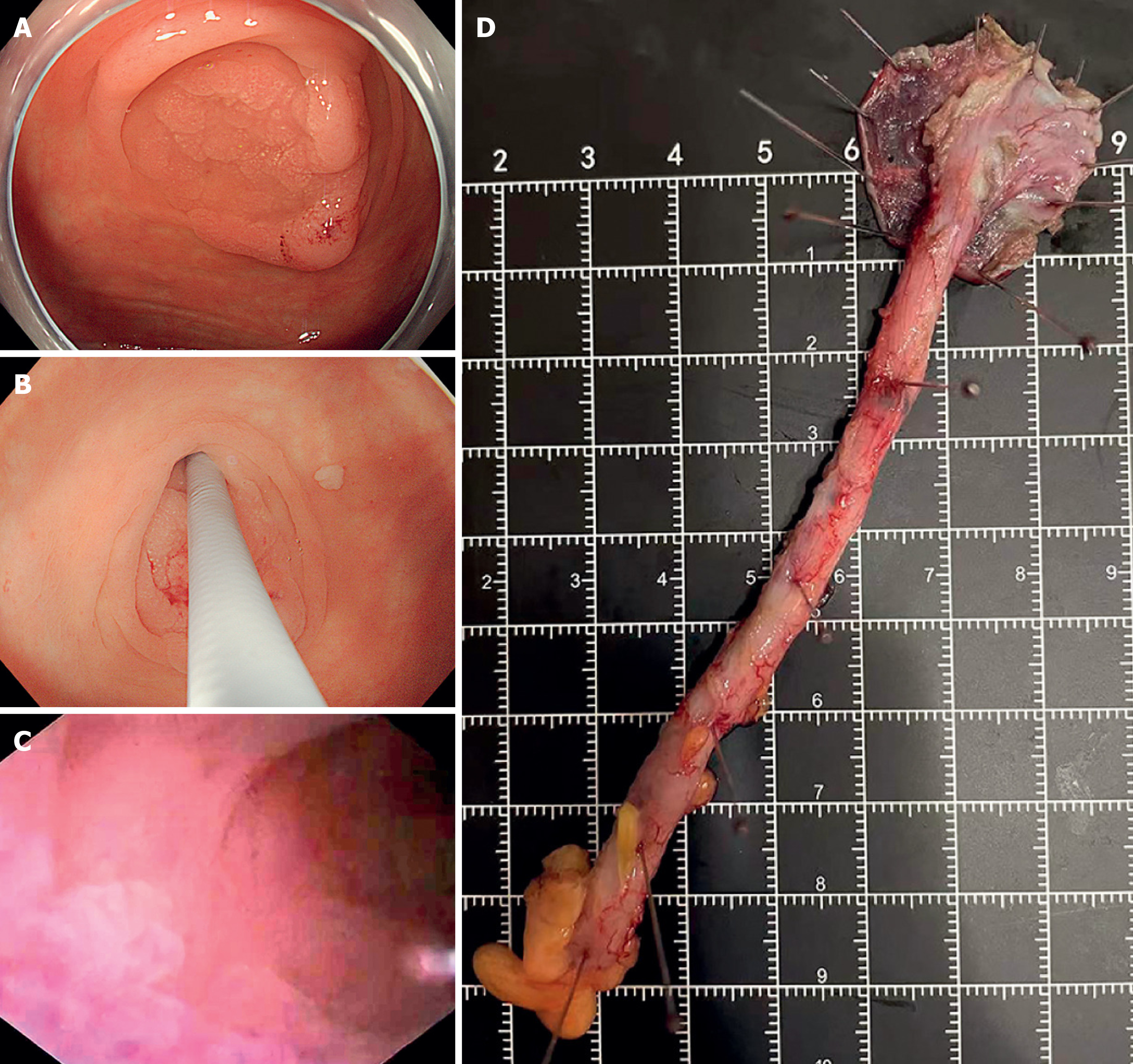
Figure 2 Single-use cholangioscope-assisted diagnosis of sessile serrated lesions within the appendix[7].
A: Colonoscopy revealed a 20-mm laterally spreading tumor in the ileocecal region; B: A cholangioscope was utilized to further examine the appendix; C: The rough, granular mucosa was unveiled within the appendix cavity; D: Removed appendix. Citation: Yao J, Liu K, Zhao G, Wang Z, Wang X, Fu J. Endoscopic management of multiple sessile serrated lesions in both the ileocecal region and the appendix cavity. Endoscopy 2024; 56: E841-E842. Copyright ©The Author(s) 2024. Published by Thieme (Supplementary material).
- Citation: Zhang MY, Yao JJ, Pan SX, Hou WW, Wei X, Zhao XL, Fu JD. Sessile serrated lesions involving the appendiceal orifice: Endoscopic diagnosis and treatment. World J Gastrointest Endosc 2025; 17(10): 110417
- URL: https://www.wjgnet.com/1948-5190/full/v17/i10/110417.htm
- DOI: https://dx.doi.org/10.4253/wjge.v17.i10.110417