
















©The Author(s) 2025.
World J Gastrointest Endosc. Oct 16, 2025; 17(10): 110417
Published online Oct 16, 2025. doi: 10.4253/wjge.v17.i10.110417
Published online Oct 16, 2025. doi: 10.4253/wjge.v17.i10.110417
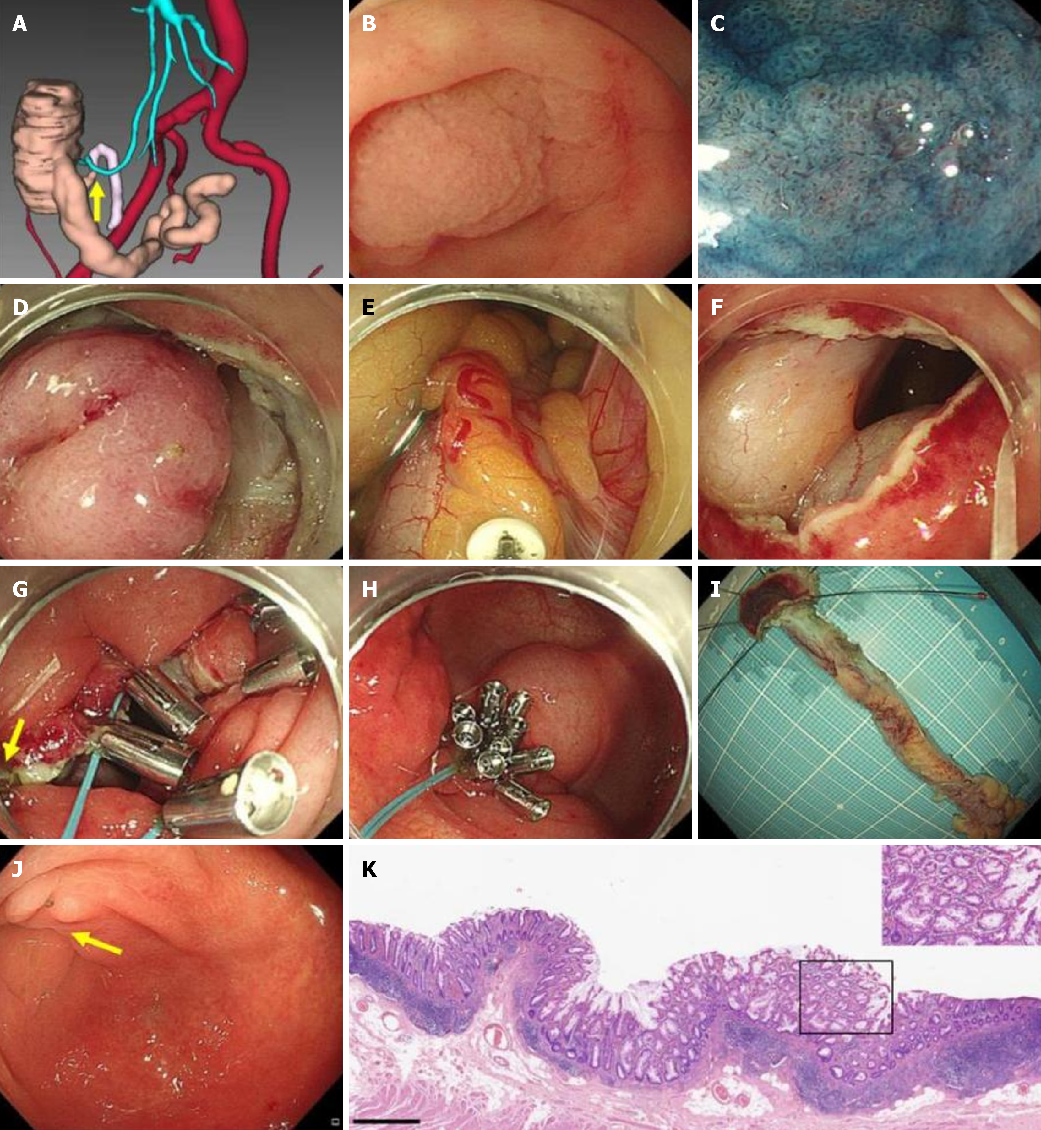
Figure 1 Transcolonic endoscopic appendectomy for the sessile serrated lesion involving the appendiceal orifice[35].
A: Three-dimensional reconstruction images showing the appendix (yellow arrow) and adjacent bowels and vessels; B: Endoscopic white-light image showing the sessile serrated lesion (SSL) involving the appendiceal orifice (AO); C: Image from chromoendoscopy following indigo carmine dye spraying clearly showing the SSL; D: Intraprocedural view showing endoscopic full-thickness resection of the cecum tissue around the AO; E: Endoscopic dissection of the mesoappendix along the appendix by an insulated-tipped knife; F: Cecal defection; G: Intraprocedural endoscopic image showing clips and endoloop used for closing the cecal defect (yellow arrow: The dental-floss assistance); H: Cecal defect was perfectly closed by clips and endoloop after transcolonic endoscopic appendectomy; I: The specimen was calculated and examined; J: Endoscopic follow-up image showing the cecum 3 months after discharge (yellow arrow: The wound healing scar); K: Pathological confirmation and diagnosis of the SSL (bar: 100 µm): Increased gland diameter and enlarged opening; microbubble-like mucous cells; jagged crypts, widened and inverted crypt base. Citation: Chen T, Xu A, Lian J, Chu Y, Zhang H, Xu M. Transcolonic endoscopic appendectomy: A novel natural orifice transluminal endoscopic surgery (NOTES) technique for the sessile serrated lesions involving the appendiceal orifice. Gut 2021; 70: 1812-1814. Copyright ©The Author(s) 2021. Published by BMJ (Supplementary material).
- Citation: Zhang MY, Yao JJ, Pan SX, Hou WW, Wei X, Zhao XL, Fu JD. Sessile serrated lesions involving the appendiceal orifice: Endoscopic diagnosis and treatment. World J Gastrointest Endosc 2025; 17(10): 110417
- URL: https://www.wjgnet.com/1948-5190/full/v17/i10/110417.htm
- DOI: https://dx.doi.org/10.4253/wjge.v17.i10.110417