
















©The Author(s) 2025.
World J Gastrointest Endosc. Oct 16, 2025; 17(10): 108729
Published online Oct 16, 2025. doi: 10.4253/wjge.v17.i10.108729
Published online Oct 16, 2025. doi: 10.4253/wjge.v17.i10.108729
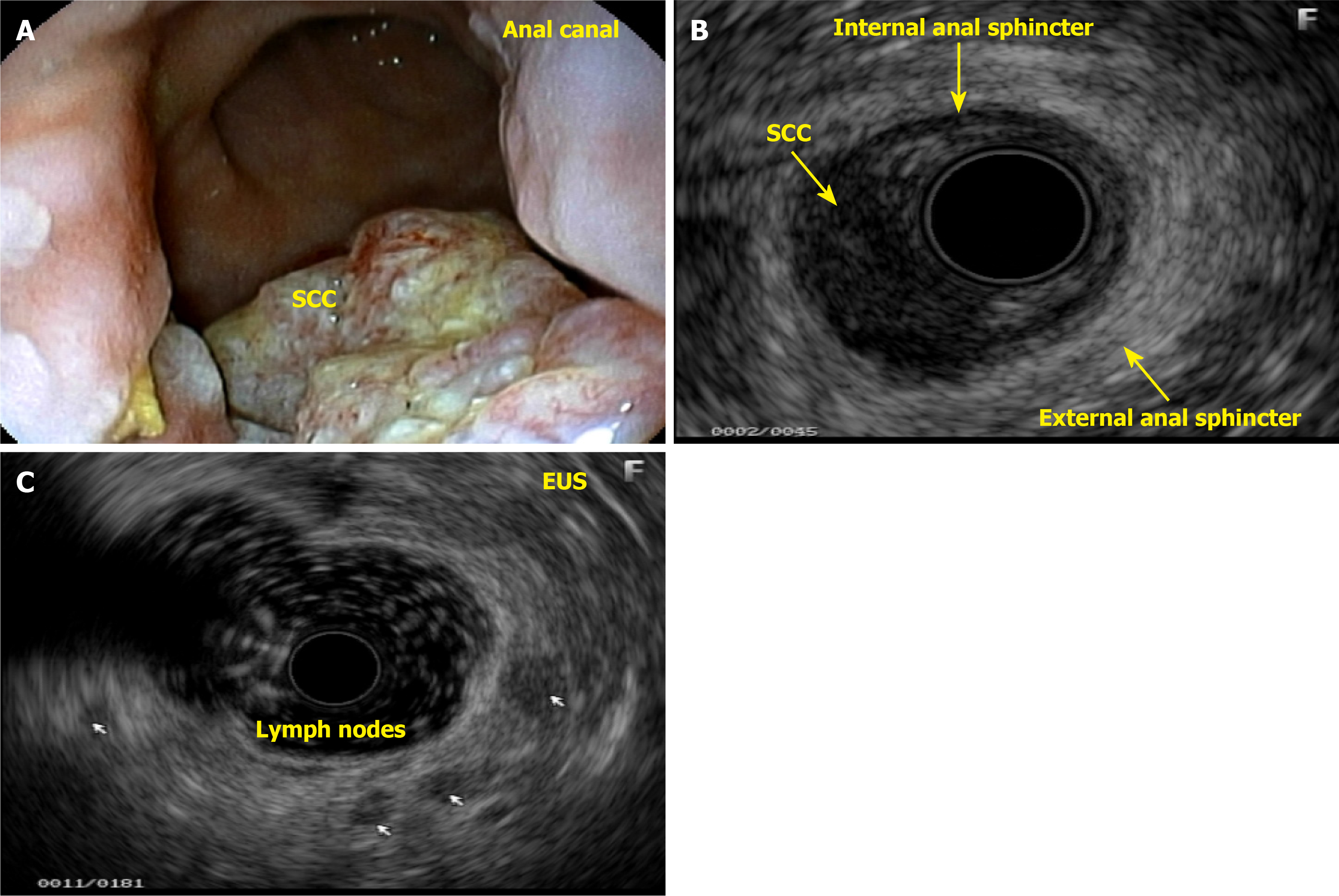
Figure 11 Squamous cell carcinoma in the anal canal.
A: Ulcerative-vegetative lesion measuring approximately 3 cm in length; B: Endoscopic ultrasound showing a hypoechoic, irregular, and heterogeneous lesion extending to the anterior wall of the lower rectum and infiltrating the internal and external anal sphincters, as well as the perirectal fat, affecting approximately 30% of the circumference of the wall, anterior face; C: Presence of 5 hypoechoic, regular, and homogeneous lymph nodes, some larger than 1 cm, bilateral, inguinal, and perirectal (-uT2N3Mx). SCC: Squamous cell carcinoma; EUS: Endoscopic ultrasound.
- Citation: Gomes A, Bara J, da Matta CHKT, Paiva PP, de Carvalho LM, Bononi BM, Pinto PCC, Rodrigues JMS, Borghesi RA. Anal neoplasm in colonoscopy: What endoscopists need to know. World J Gastrointest Endosc 2025; 17(10): 108729
- URL: https://www.wjgnet.com/1948-5190/full/v17/i10/108729.htm
- DOI: https://dx.doi.org/10.4253/wjge.v17.i10.108729