
















©The Author(s) 2025.
World J Gastrointest Endosc. Oct 16, 2025; 17(10): 107792
Published online Oct 16, 2025. doi: 10.4253/wjge.v17.i10.107792
Published online Oct 16, 2025. doi: 10.4253/wjge.v17.i10.107792
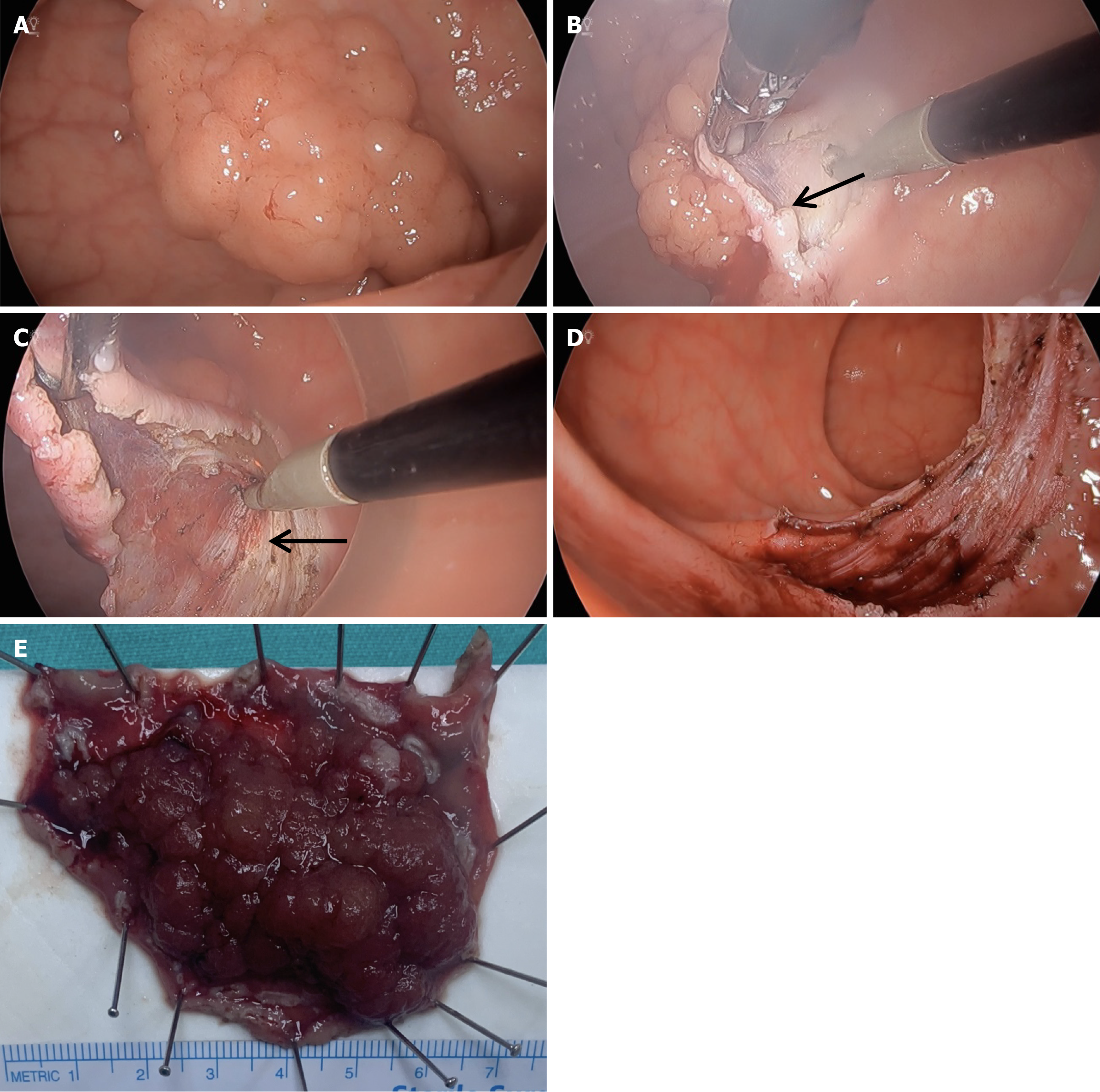
Figure 2 Steps of transanal endoscopic microsurgery-assisted endoscopic submucosal dissection for a rectal lesion.
A: Endoscopic view of the nodular lesion; B: Submucosal dissection is initiated (black arrow); C: Muscularis propria layer is preserved during dissection (black arrow); D: Post-dissection view showing the complete resection site with intact muscularis propria; E: Resected lesion pinned on a board for pathological assessment.
- Citation: Ilhan E, Cengiz F. Endoscopic submucosal dissection, transanal endoscopic microsurgical submucosal dissection, and transanal minimally invasive surgery in rectal lesions. World J Gastrointest Endosc 2025; 17(10): 107792
- URL: https://www.wjgnet.com/1948-5190/full/v17/i10/107792.htm
- DOI: https://dx.doi.org/10.4253/wjge.v17.i10.107792