
















©The Author(s) 2025.
World J Gastrointest Endosc. Jan 16, 2025; 17(1): 101119
Published online Jan 16, 2025. doi: 10.4253/wjge.v17.i1.101119
Published online Jan 16, 2025. doi: 10.4253/wjge.v17.i1.101119
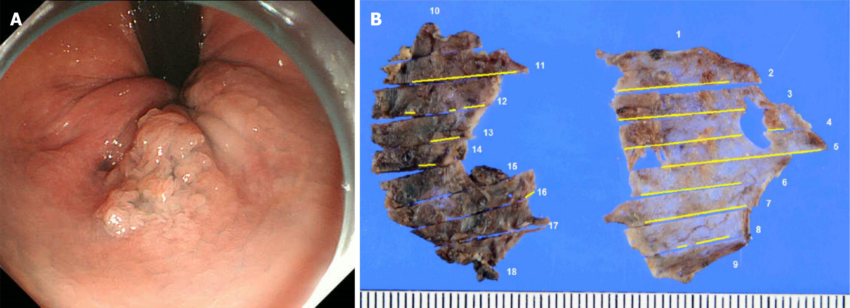
Figure 1 Endoscopic image at initial presentation.
A: A 20-mm elevated lesion on the left wall of the anal canal; B: The number represents the cross section, and the tumor is identified by the yellow line. But the specimen was removed in two sections. Therefore, it was difficult to evaluate.
- Citation: Kinoshita M, Maruyama T, Hike S, Hirosuna T, Kainuma S, Kinoshita K, Nakano A, Ohira G, Uesato M, Matsubara H. Complete resection of recurrent anal canal cancer using endoscopic submucosal dissection and transanal resection: A case report. World J Gastrointest Endosc 2025; 17(1): 101119
- URL: https://www.wjgnet.com/1948-5190/full/v17/i1/101119.htm
- DOI: https://dx.doi.org/10.4253/wjge.v17.i1.101119