
















©The Author(s) 2024.
World J Gastrointest Endosc. Aug 16, 2024; 16(8): 494-499
Published online Aug 16, 2024. doi: 10.4253/wjge.v16.i8.494
Published online Aug 16, 2024. doi: 10.4253/wjge.v16.i8.494
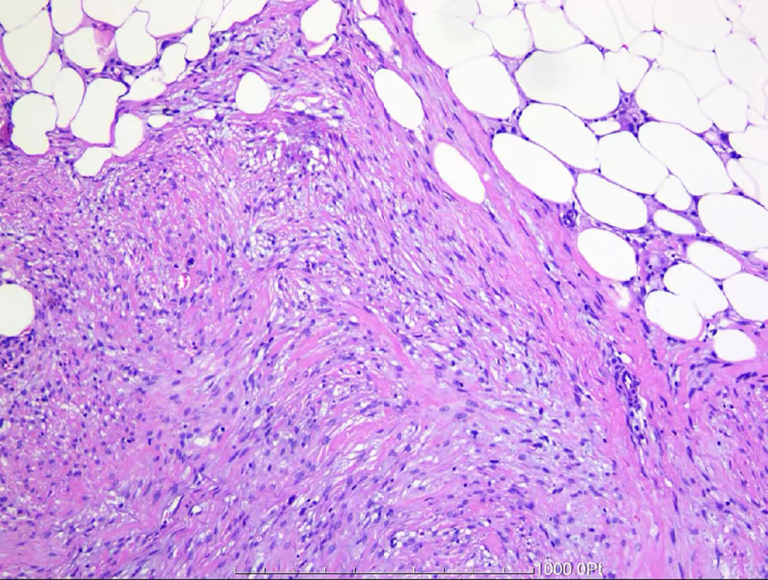
Figure 3 Postoperative pathological examination of tissue specimens.
Surgical pathology findings revealed spindle-shaped cells and adipocytes distributed in a patchy pattern within the tough area of the outer mesentery of the colonic wall, with spindle cells arranged in bundles or interlaced (some in a disordered manner). The cells had short spindle-shaped nuclei, which were slightly pointed at both ends, and stained cytoplasm. These findings suggested focal new bone formation, which was diagnosed as heterotopic mesenteric ossification.
- Citation: Zhang BF, Liu J, Zhang S, Chen L, Lu JZ, Zhang MQ. Heterotopic mesenteric ossification caused by trauma: A case report. World J Gastrointest Endosc 2024; 16(8): 494-499
- URL: https://www.wjgnet.com/1948-5190/full/v16/i8/494.htm
- DOI: https://dx.doi.org/10.4253/wjge.v16.i8.494