Copyright
©The Author(s) 2024.
World J Gastrointest Endosc. Mar 16, 2024; 16(3): 136-147
Published online Mar 16, 2024. doi: 10.4253/wjge.v16.i3.136
Published online Mar 16, 2024. doi: 10.4253/wjge.v16.i3.136
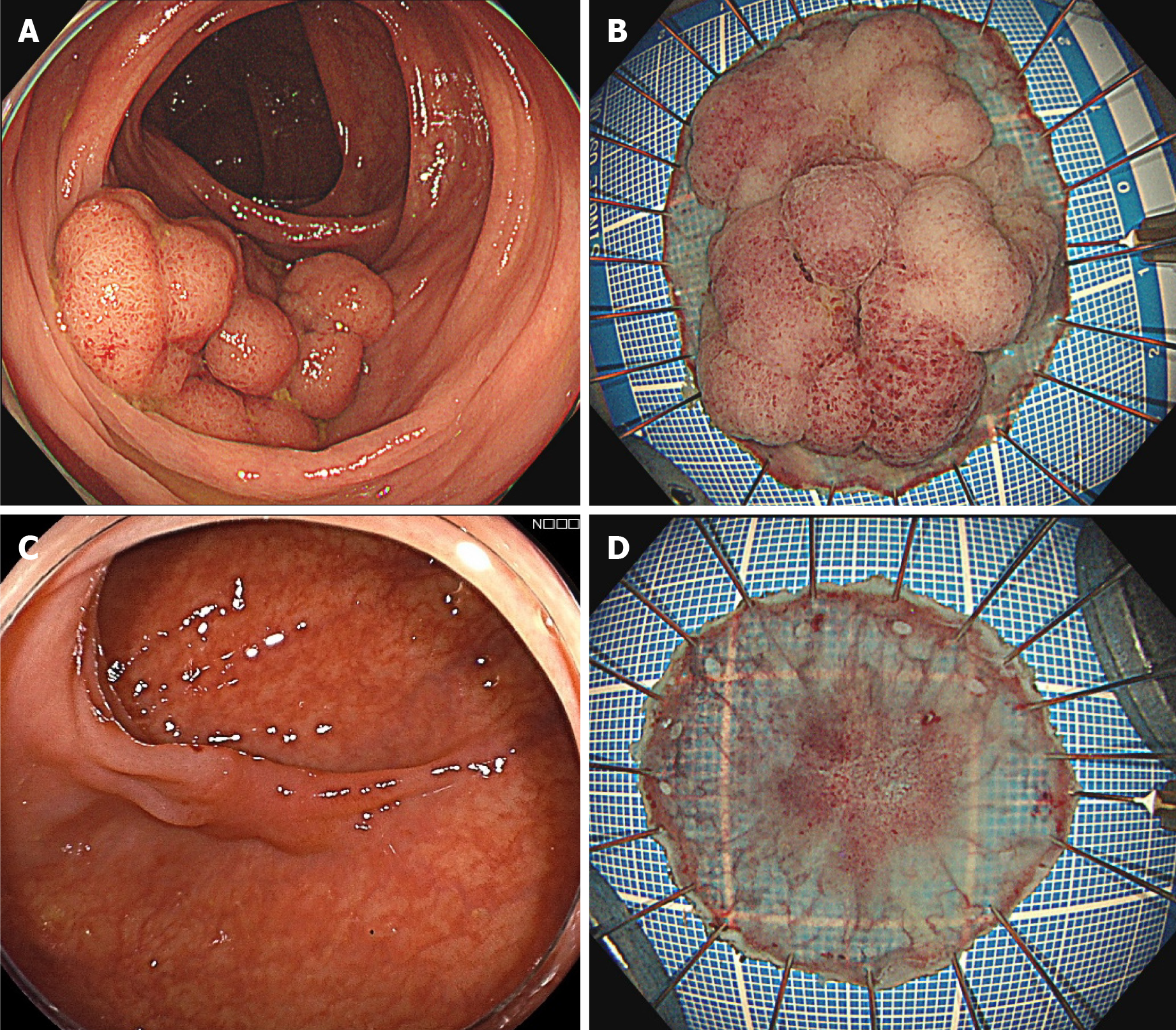
Figure 2 Representative cases of incorrect scaling.
A: Underscaling case. The tumor size was evaluated as 30 mm; B: Pathology revealing the maximal diameter as 49 mm, wherein the size discrepancy was -39%; C: Overscaling case. The cancerous lesion was evaluated as 20 mm; D: The pathological size was 15 mm, resulting in a size discrepancy of 33%.
- Citation: Onda T, Goto O, Otsuka T, Hayasaka Y, Nakagome S, Habu T, Ishikawa Y, Kirita K, Koizumi E, Noda H, Higuchi K, Omori J, Akimoto N, Iwakiri K. Tumor size discrepancy between endoscopic and pathological evaluations in colorectal endoscopic submucosal dissection. World J Gastrointest Endosc 2024; 16(3): 136-147
- URL: https://www.wjgnet.com/1948-5190/full/v16/i3/136.htm
- DOI: https://dx.doi.org/10.4253/wjge.v16.i3.136


