Copyright
©The Author(s) 2023.
World J Gastrointest Endosc. Mar 16, 2023; 15(3): 84-102
Published online Mar 16, 2023. doi: 10.4253/wjge.v15.i3.84
Published online Mar 16, 2023. doi: 10.4253/wjge.v15.i3.84
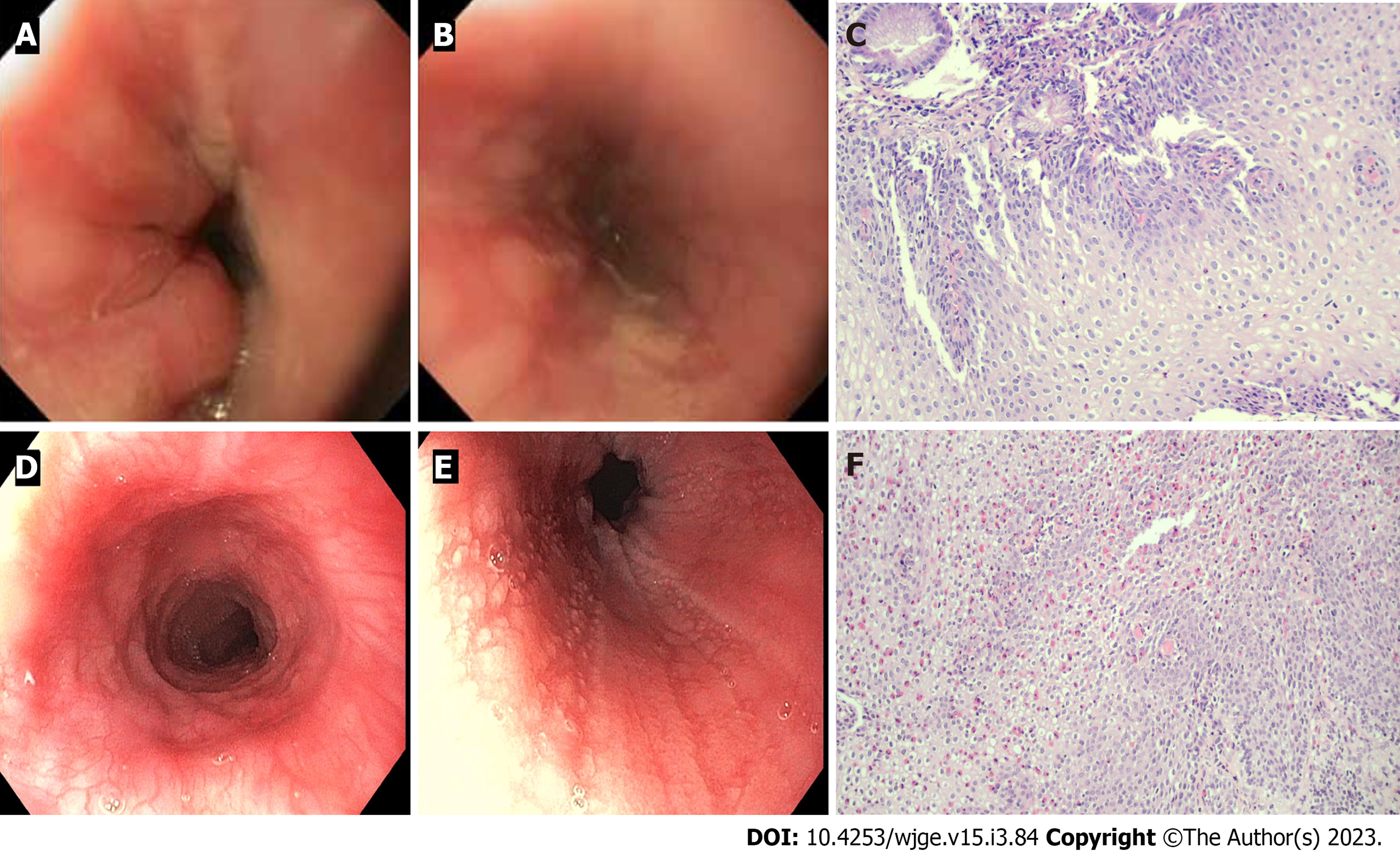
Figure 4 The endoscopic and histologic findings of reflux esophagitis and eosinophilic esophagitis.
A and B : Endoscopic finding of reflux esophagitis shows mucosal breaks and healing mucosal damage; C: Histopathology section (× 20) showing basal cell hyperplasia, elongation of the lamina propria papillae and scattered eosinophilic infiltration; D and E: Endoscopic findings of eosinophilic esophagitis showing ringed esophagus, linear furrows and whitish papules; F: Histopathology section (× 20) showing numerous eosinophils diffusely infiltrating the squamous epithelium (peak eosinophilic count = 40 cells/HPF). The squamous epithelium reveals spongiosis. Eosinophilic microabscesses and eosinophil degranulation are also noted.
- Citation: Sintusek P, Mutalib M, Thapar N. Gastroesophageal reflux disease in children: What’s new right now? World J Gastrointest Endosc 2023; 15(3): 84-102
- URL: https://www.wjgnet.com/1948-5190/full/v15/i3/84.htm
- DOI: https://dx.doi.org/10.4253/wjge.v15.i3.84


