Copyright
©The Author(s) 2022.
World J Gastrointest Endosc. Sep 16, 2022; 14(9): 536-546
Published online Sep 16, 2022. doi: 10.4253/wjge.v14.i9.536
Published online Sep 16, 2022. doi: 10.4253/wjge.v14.i9.536
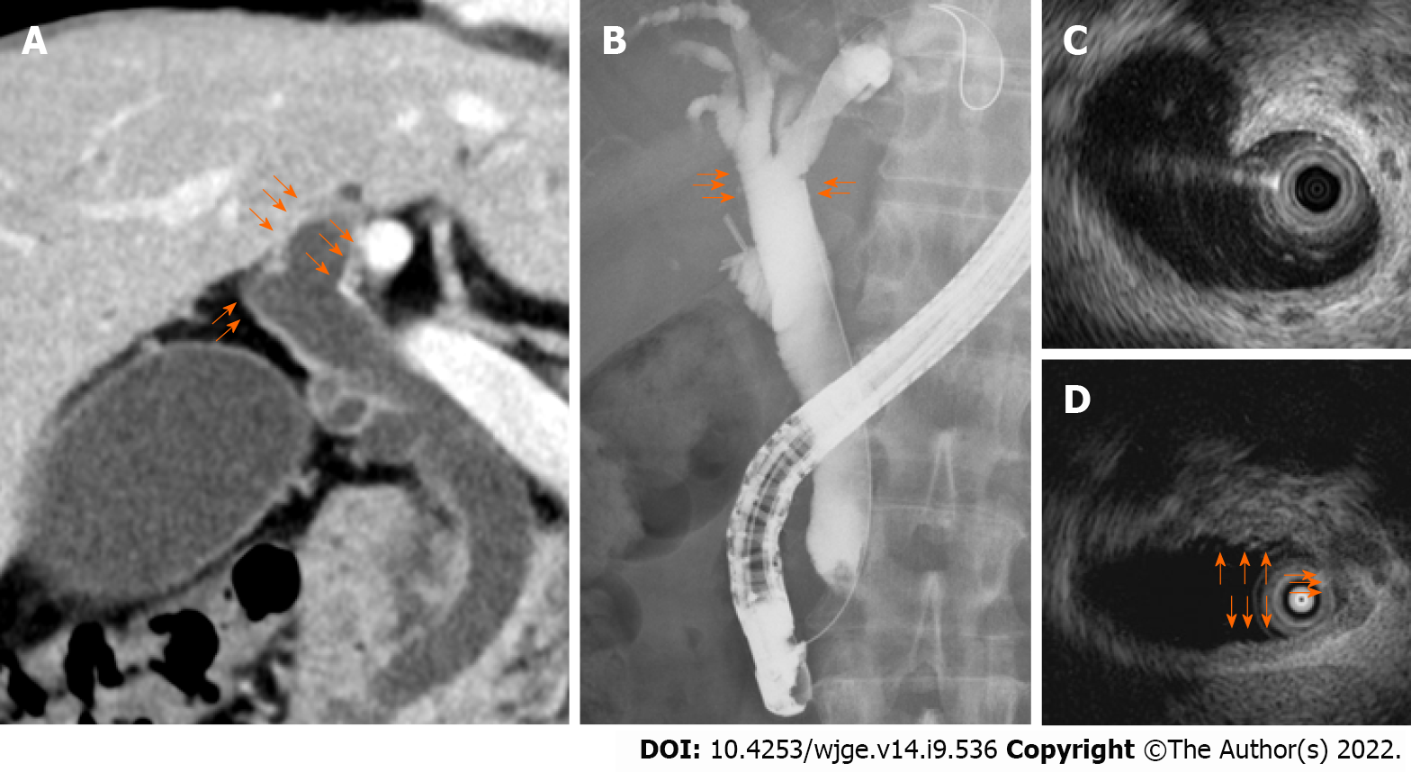
Figure 1 Imaging findings of the hilar biliary duct.
A: Thickening and enhancement of the bile duct wall on contrast-enhanced computed tomography; B: Irregularity on endoscopic retrograde cholangiography; C: Thickening of the entire bile duct wall on intraductal ultrasonography (IDUS); D: Partial thickening of the bile duct wall on IDUS.
- Citation: Takagi T, Sugimoto M, Suzuki R, Konno N, Asama H, Sato Y, Irie H, Nakamura J, Takasumi M, Hashimoto M, Kato T, Kobashi R, Yanagita T, Hashimoto Y, Marubashi S, Hikichi T, Ohira H. Screening for hilar biliary invasion in ampullary cancer patients. World J Gastrointest Endosc 2022; 14(9): 536-546
- URL: https://www.wjgnet.com/1948-5190/full/v14/i9/536.htm
- DOI: https://dx.doi.org/10.4253/wjge.v14.i9.536


